
Vertebral column (spine)
Columna vertebralis
The vertebral column, or spine, is the stack of bones that runs down the middle of the back from the skull to the pelvis. It has 24 movable vertebrae in three regions, followed by the sacrum and the coccyx.
Loading 3D model…
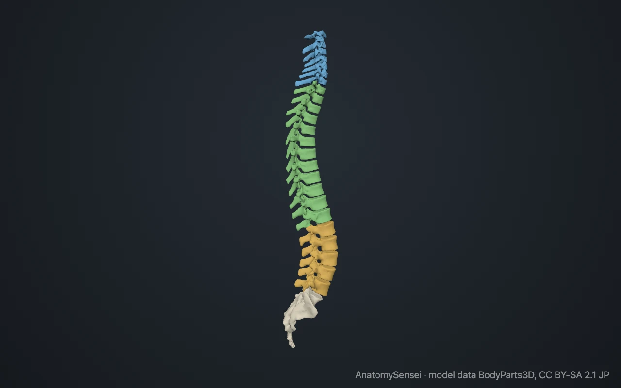
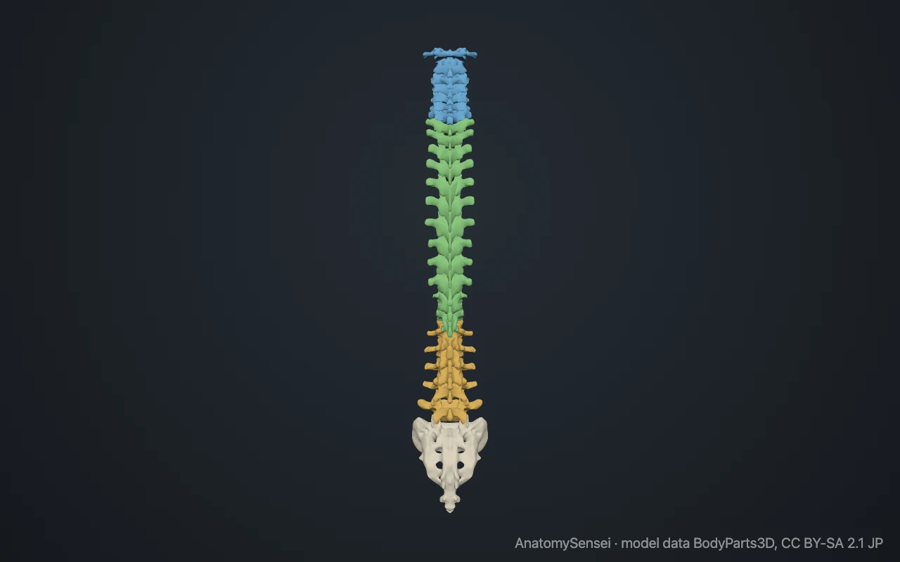
Free 3D model of the vertebrae. Whole spine. Drag to rotate, zoom with + and − (or Ctrl + scroll), click a bone to highlight and name it.
- Cervical vertebrae (C1 to C7)
- Thoracic vertebrae (T1 to T12)
- Lumbar vertebrae (L1 to L5)
Key points
- The spine has 24 separate vertebrae: 7 cervical, 12 thoracic and 5 lumbar.
- Below them, five fused vertebrae form the sacrum, and the small coccyx ends the column.
- Seen from the side, the spine has four gentle curves that help it carry load.
- A typical vertebra has a body in front and an arch behind, with the spinal cord running between them.
What is the vertebral column?
The vertebral column is the central pillar of the skeleton. It is built from a chain of bones called vertebrae, stacked one on top of the next from the base of the skull to the tailbone. Everyday names for it are the spine and the backbone.
An adult usually has 33 vertebrae in all. The upper 24 stay separate and can move on each other. The lower ones fuse during growth into two bones, the sacrum and the coccyx.
The model on this page shows the 24 separate vertebrae, each as its own bone, with the sacrum beneath them. It shows bones only, so the discs between the vertebrae appear as gaps, and the coccyx is not included.
What are the regions of the spine?
The movable part of the spine is divided into three regions. The seven cervical vertebrae (C1 to C7) form the neck. The twelve thoracic vertebrae (T1 to T12) form the upper and middle back, and each carries a pair of ribs. The five lumbar vertebrae (L1 to L5) form the lower back.
Vertebrae are numbered from the top down within each region. The first two, the atlas (C1) and the axis (C2), have their own names because their shapes are unlike any others.
Below L5 comes the sacrum, a triangular bone made of five fused vertebrae that is wedged between the two hip bones. The coccyx, or tailbone, is the small final segment, usually made of four tiny fused vertebrae.
Why is the spine curved?
Seen from the front or the back, a healthy spine is straight. Seen from the side, it has four gentle curves. The neck and the lower back curve forward, a shape called lordosis. The thoracic region and the sacrum curve backward, a shape called kyphosis.
The thoracic and sacral curves are present before birth. The cervical and lumbar curves develop as a baby learns to hold up the head and then to stand and walk. Together the curves let the spine act like a spring, which absorbs shock better than a straight column would and keeps the head balanced over the pelvis.
What are the parts of a typical vertebra?
Most vertebrae share the same plan. In front is the vertebral body, a short cylinder of bone that carries weight. Behind it is the vertebral arch, a ring of bone made of two pedicles and two flat plates called laminae.
The body and the arch enclose a hole, the vertebral foramen. Stacked together, these holes form the vertebral canal, the bony tunnel that holds the spinal cord.
Seven projections stand out from the arch. One spinous process points backward in the midline, and the row of them forms the ridge that can be felt down the back. Two transverse processes point out to the sides. Four articular processes, two above and two below, link each vertebra to its neighbours.
How are the vertebrae joined together?
Neighbouring vertebrae meet in three places. In front, the two bodies are joined by an intervertebral disc, a tough pad with a firm outer ring and a soft, gel-like centre. The discs cushion load and allow a little movement in every direction.
Behind, a pair of small facet joints links the articular processes of one vertebra to those of the next. The angle of these joints differs between regions, which is why the neck turns freely while the lower back mostly bends forward and back.
There are 23 discs. None lies between the skull and the atlas or between the atlas and the axis. Between each pair of vertebrae, an opening on each side lets a spinal nerve leave the canal.
What does the spine connect to?
At the top, the atlas carries the skull by meeting the occipital bone. In the chest, the twelve thoracic vertebrae join the twelve pairs of ribs. At the bottom, L5 sits on the sacrum, and the sacrum joins the two hip bones at the sacroiliac joints. In this way body weight passes from the spine into the pelvis and on to the legs.
What does the spine do?
The spine has four main jobs. It supports the head and trunk and passes their weight down to the pelvis. It protects the spinal cord inside the vertebral canal. It allows the trunk to bend and twist. It also gives attachment to the ribs and to the many muscles and ligaments of the back.
Each joint between two vertebrae moves only a little. The large range of the whole spine comes from adding these small movements together.
Common problems with the spine
Back and neck pain are among the most common complaints in medicine, and most often involve the mobile cervical and lumbar regions. A herniated disc occurs when the soft centre of a disc bulges through its outer ring, most often low in the lumbar spine. Wear of the discs and facet joints with age is called spondylosis.
Scoliosis is a sideways curve of the spine. An increased backward curve of the thoracic region is called hyperkyphosis. Vertebral bodies weakened by osteoporosis can collapse in a compression fracture, typically in the thoracic and upper lumbar spine. This page describes anatomy only.
Free 3D models of the vertebrae
Common questions
How many bones are in the spine?
An adult has 24 separate vertebrae, plus the sacrum and the coccyx. Counting the fused segments individually gives 33 vertebrae in most people.
How many vertebrae are in each region?
Seven cervical, twelve thoracic and five lumbar. The sacrum is made of five fused vertebrae and the coccyx usually of four.
Is the spinal cord as long as the spine?
No. In adults the cord usually ends at about the level of the first or second lumbar vertebra. Below that the canal holds a bundle of nerve roots.
What is the difference between the spine and the spinal cord?
The spine is the column of bones. The spinal cord is the nerve tissue that runs inside it, through the vertebral canal.
Is this 3D model of the vertebrae free?
Yes. The interactive 3D model on this page is free to view and use for study, with no account or download needed. Its geometry comes from the open BodyParts3D data set (CC BY-SA).
About this free 3D model of the vertebrae
The interactive 3D model on this page is free to use in your browser, with no sign-up. It is built from scan-based anatomy data, so every bone is a separate, correctly placed shape you can turn and inspect from any side.