
Lumbar vertebrae
Vertebrae lumbales
The lumbar vertebrae are the five bones of the lower back, numbered L1 to L5. They are the largest and strongest of the movable vertebrae because they carry the most body weight.
Loading 3D model…
Free 3D model of the lumbar vertebrae. Whole spine. Drag to rotate, zoom with + and − (or Ctrl + scroll), click a bone to highlight and name it.
Key points
- There are five lumbar vertebrae, L1 to L5, between the rib cage and the pelvis.
- They have the largest bodies of any vertebrae because they bear the most weight.
- Their short, thick spinous processes point straight back.
- L5 rests on the sacrum at the lumbosacral joint, a common site of back problems.
What are the lumbar vertebrae?
The lumbar vertebrae are the five vertebrae of the lower back. They form the last region of the movable spine, below the thoracic vertebrae and above the sacrum.
Each level carries the weight of everything above it, so the lumbar vertebrae are the biggest in the body. They have no ribs attached, which leaves the lower back free to bend.
Where are the lumbar vertebrae?
They lie between the bottom of the rib cage and the top of the pelvis. Seen from the side they form a forward curve, the lumbar lordosis, which gives the lower back its hollow.
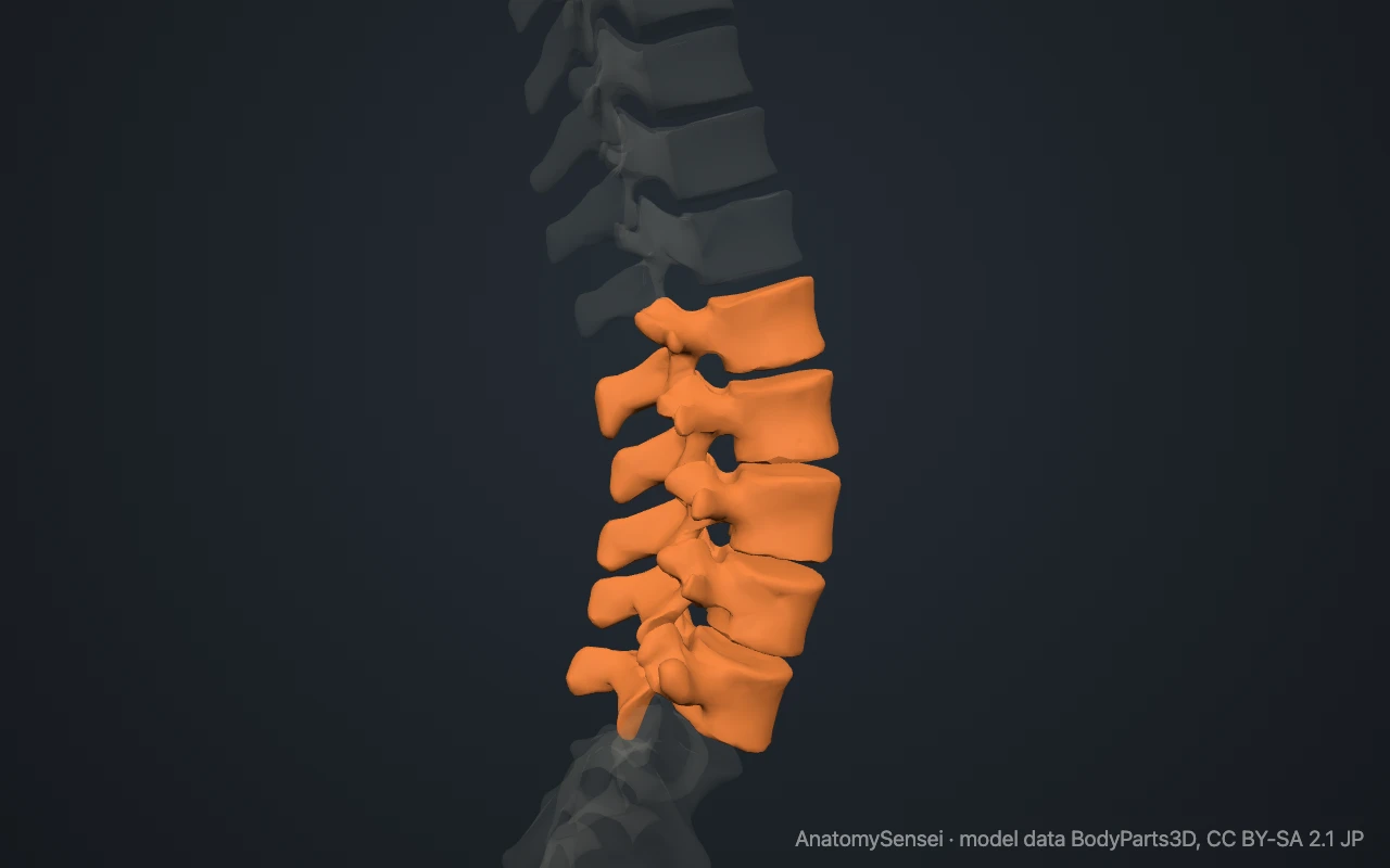
You can find them on your own back. Place your hands on your hips and feel for the top edges of the hip bones. A line joining those two points across the back crosses the spine at about L4. The spinous processes above and below can be felt in the midline.
What makes a lumbar vertebra different?
A lumbar vertebra is recognised by what it lacks as much as by what it has. It has no facets for ribs and no holes in its transverse processes. Its body is large, wider from side to side than from front to back, and kidney-shaped from above.
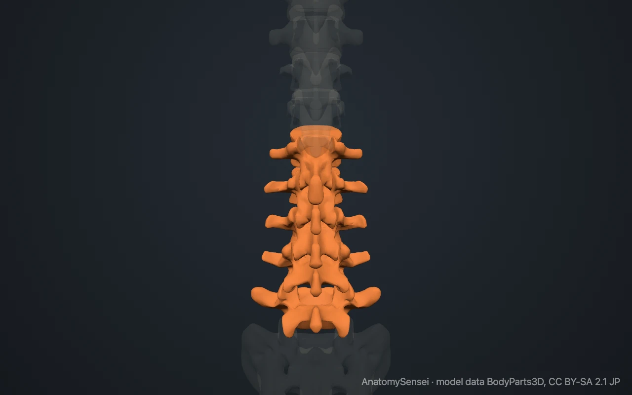
The spinous process is short, thick and squared, like the blade of a hatchet, and points straight backward. The transverse processes are long and slender. The vertebral foramen is triangular.
The facet joints face mostly inward and outward. This arrangement lets the lower back bend forward and backward freely but allows very little twisting.
How do L1 and L5 differ from the others?
L5 is the largest vertebra and the most distinctive of the five. Its body is taller in front than behind, which makes it wedge-shaped, and its transverse processes are short and stout. Strong ligaments tie them to the pelvis.
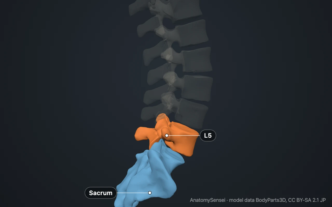
L5 sits on the sloping top of the sacrum at the lumbosacral joint. Here the forward curve of the lumbar spine turns sharply into the backward curve of the sacrum, so the joint is under considerable stress.
At the upper end, L1 marks roughly where the spinal cord ends in adults. Below that level the vertebral canal contains a bundle of nerve roots called the cauda equina, the "horse's tail".
What do the lumbar vertebrae articulate with?
L1 joins T12 above, and L5 joins the sacrum below. Between neighbours there is a disc in front and a pair of facet joints behind, as elsewhere in the spine. The lumbar discs are the thickest in the column.
What attaches to the lumbar vertebrae?
The psoas major muscle arises from the sides of the lumbar bodies and transverse processes and runs down to the femur, where it bends the hip. The two crura of the diaphragm attach to the front of the upper lumbar bodies.
Behind, the erector spinae and the deeper multifidus muscles attach to the spinous and transverse processes. The quadratus lumborum connects the transverse processes to the twelfth rib and the hip bone.
What do the lumbar vertebrae do?
They carry the weight of the upper body and pass it to the sacrum and pelvis. They also provide most of the forward and backward bending of the trunk, and some bending to the side.
The canal inside them protects the lower end of the spinal cord and the nerve roots that go on to the pelvis and legs.
Common problems with the lumbar vertebrae
Low back pain most often arises in this region. Disc herniation is most frequent at the two lowest levels, between L4 and L5 and between L5 and the sacrum. A herniated disc there can press on a nerve root and cause pain down the leg, known as sciatica.
A stress fracture of the arch of a vertebra, most often L5, is called spondylolysis. If the body then slips forward on the one below, the condition is spondylolisthesis. Narrowing of the canal with age is called lumbar spinal stenosis. This page describes anatomy only.
Common questions
How many lumbar vertebrae are there?
Five in most people, numbered L1 to L5. A few people have four or six because the lowest one is fused with the sacrum or the first sacral segment is separate.
Which is the largest vertebra in the spine?
L5, the lowest lumbar vertebra, which carries the most weight.
Why is a lumbar puncture done in the lower back?
The spinal cord usually ends near L1 or L2, so a needle placed between the lower lumbar vertebrae enters the canal below the cord.
Do the lumbar vertebrae have ribs?
No. Their long transverse processes take the place of ribs at these levels.
Is this 3D model of the lumbar vertebrae free?
Yes. The interactive 3D model on this page is free to view and use for study, with no account or download needed. Its geometry comes from the open BodyParts3D data set (CC BY-SA).
About this free 3D model of the lumbar vertebrae
The interactive 3D model on this page is free to use in your browser, with no sign-up. It is built from scan-based anatomy data, so every bone is a separate, correctly placed shape you can turn and inspect from any side.