
Occipital bone
Os occipitale
The occipital bone is the single bone at the back and base of the skull. It surrounds the foramen magnum, the large opening for the spinal cord, and rests on the first neck vertebra.
Loading 3D model…
Free 3D model of the occipital bone. Whole skull. Drag to rotate, zoom with + and − (or Ctrl + scroll), click a bone to highlight and name it.
Key points
- The occipital bone forms the back of the head and part of the skull base.
- The foramen magnum in its base is where the brain continues into the spinal cord.
- Two occipital condyles rest on the atlas and let the head nod.
- The bump at the back of the head is its external occipital protuberance.
What is the occipital bone?
The occipital bone is the cranial bone at the back and underside of the skull. It is shaped like a shallow dish, curved both from side to side and from top to bottom.
It is the only skull bone that forms a joint with the spine. It supports the back of the brain, including the cerebellum.
Where is the occipital bone?
It lies at the lower back of the head, below the two parietal bones and between the two temporal bones. From there it curves forward under the skull to meet the sphenoid.
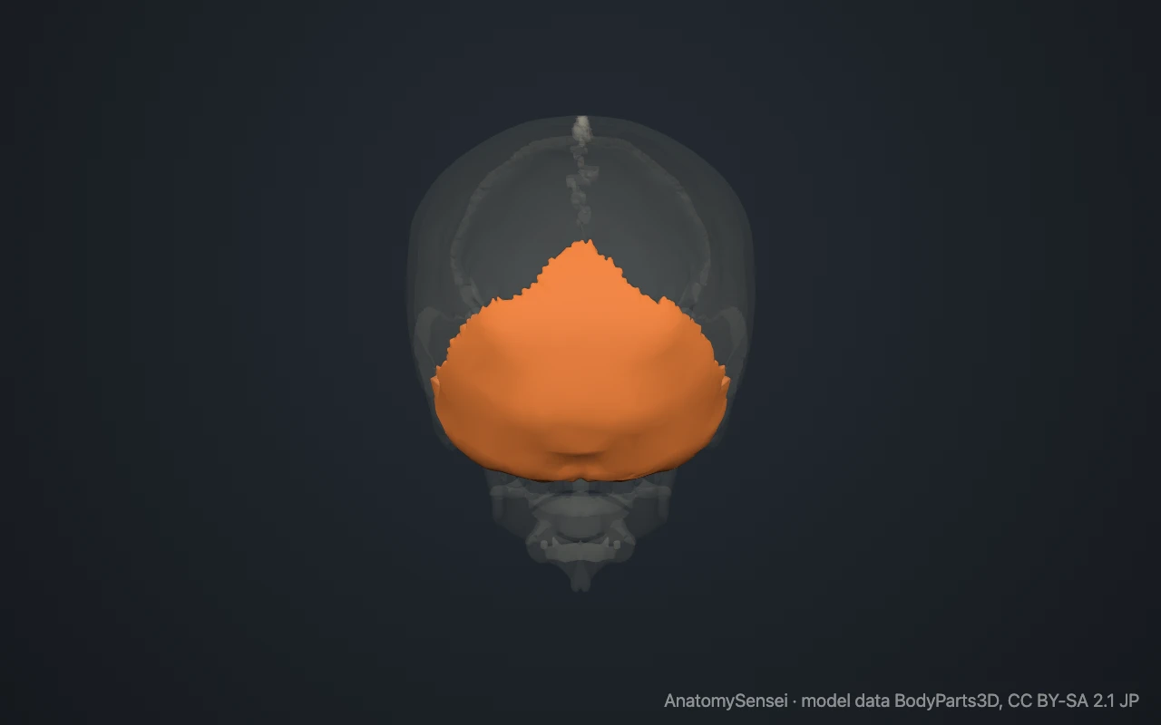
You can feel it by running a finger down the midline of the back of the head. The firm bump met just above the hollow of the neck is the external occipital protuberance. The neck muscles hide the bone below this point.
What are the parts of the occipital bone?
The bone is arranged around the foramen magnum, a large oval opening. The broad plate behind the opening is the squamous part. The thick bar in front of it is the basilar part, and the two lateral parts lie at its sides.
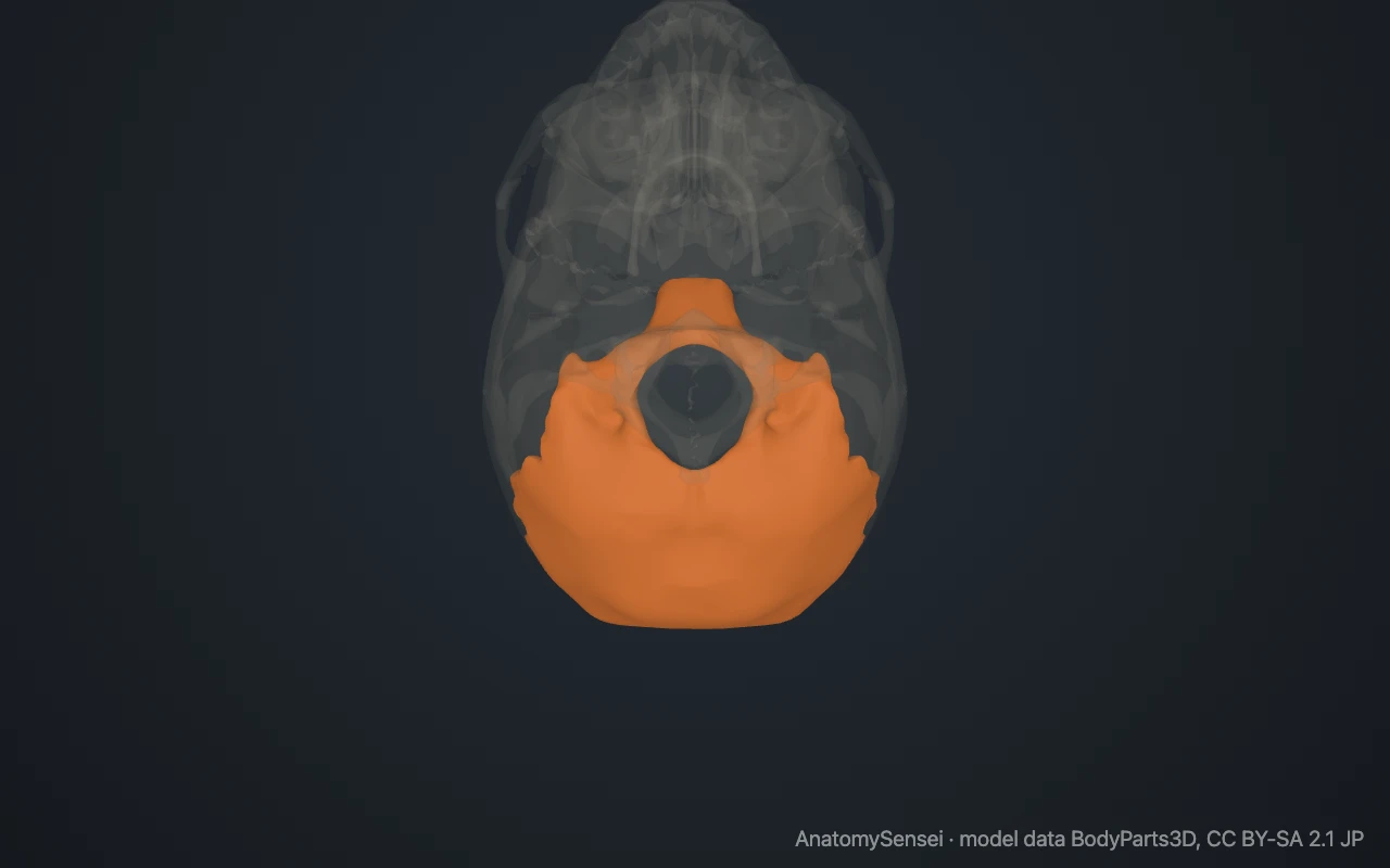
On the underside of each lateral part is an occipital condyle, a smooth, kidney-shaped knob. Above each condyle a short tunnel, the hypoglossal canal, carries the nerve that moves the tongue.
On the outer surface of the squamous part, curved ridges called the nuchal lines run sideways from the external occipital protuberance. They mark where the neck muscles attach. These features are parts of the one bone and cannot be highlighted separately in the model.
What does the occipital bone articulate with?
It meets six bones. Above, it joins the two parietal bones along the lambdoid suture, named after its resemblance to the Greek letter lambda. At each side it joins a temporal bone.
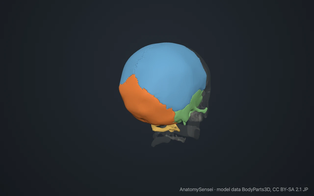
In front, its basilar part is fused to the body of the sphenoid. In a child the two are separated by a plate of cartilage, which turns to bone by early adulthood.
Below, the two occipital condyles sit in hollows on top of the atlas, the first neck vertebra. This pair of joints is the atlanto-occipital joint, and it allows the nodding movement of the head.
What attaches to the occipital bone?
Many muscles of the neck and back attach to the outer surface between and below the nuchal lines. They include the trapezius, the sternocleidomastoid, the semispinalis capitis and the small suboccipital muscles that fine-tune head position.
A strong midline ligament, the ligamentum nuchae, runs from the external occipital protuberance down the back of the neck. Inside the skull, folds of the tough membrane that covers the brain attach to ridges on the inner surface.
What does the occipital bone do?
It protects the back of the brain and provides the passage through which the brainstem continues into the spinal cord. The arteries that supply the back of the brain enter the skull through the same opening.
It also carries the weight of the head onto the spine and gives the neck muscles the leverage to hold the head upright.
Common problems with the occipital bone
A fall onto the back of the head can cause an occipital fracture. Fractures that run through the base of the skull may reach the foramen magnum or an occipital condyle.
In a Chiari malformation, the lowest part of the cerebellum extends down through the foramen magnum. This page describes anatomy only.
Common questions
What is the bump on the back of the skull?
The external occipital protuberance of the occipital bone. Its size varies and it is often more prominent in men.
What passes through the foramen magnum?
The lower end of the brainstem as it becomes the spinal cord, with its coverings, the vertebral arteries and a pair of nerves.
Which vertebra joins the occipital bone?
The atlas, the first cervical vertebra. The occipital condyles rest on it.
Is the occipital bone a cranial bone?
Yes. It is one of the eight cranial bones and one of the four that are single rather than paired.
Is this 3D model of the occipital bone free?
Yes. The interactive 3D model on this page is free to view and use for study, with no account or download needed. Its geometry comes from the open BodyParts3D data set (CC BY-SA).
About this free 3D model of the occipital bone
The interactive 3D model on this page is free to use in your browser, with no sign-up. It is built from scan-based anatomy data, so every bone is a separate, correctly placed shape you can turn and inspect from any side.