
Atlas (C1)
Atlas
The atlas is the first cervical vertebra, C1, at the very top of the spine. It is a ring of bone with no body that carries the skull and lets the head nod.
Loading 3D model…
Free 3D model of the atlas. Whole spine. Drag to rotate, zoom with + and − (or Ctrl + scroll), click a bone to highlight and name it.
Key points
- The atlas is C1, the topmost vertebra, directly under the skull.
- It is a ring with no body and no spinous process.
- Its upper joints with the occipital bone allow the head to nod.
- It turns around the dens of the axis when the head rotates.
What is the atlas?
The atlas is the first vertebra of the spine, also written C1. It is named after Atlas, the Titan of Greek myth who held up the sky, because it holds up the globe of the skull.
It looks unlike any other vertebra. It has no body and no spinous process. Instead it is a ring of bone, thicker at the sides, with a large central opening.
Where is the atlas?
It sits at the top of the neck, immediately below the skull and above the axis. It lies roughly level with the hard palate, the roof of the mouth, deep to the muscles at the back of the neck.
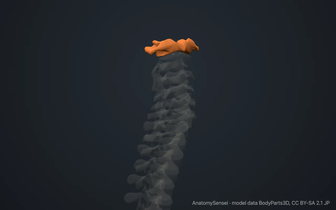
The atlas is hard to feel. Its sides reach further out than those of the vertebrae just below. With gentle pressure you may feel the tip of its transverse process in the hollow between the angle of your jaw and the bony lump behind your ear. There is no spinous process to find at the back.
What are the parts of the atlas?
The ring has four parts. On each side is a thick block of bone, the lateral mass, which carries the weight of the head. The two masses are joined in front by the short anterior arch and behind by the longer, thinner posterior arch.
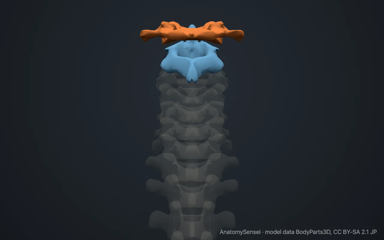
Each lateral mass has a joint surface above and below. The upper surfaces are hollow and kidney-shaped and cradle the skull. The lower ones are flatter and rest on the axis.
A transverse process projects from each lateral mass and contains a hole, the transverse foramen, for the vertebral artery. After passing through, the artery curves across a groove on top of the posterior arch on its way into the skull. A small bump, the posterior tubercle, stands where a spinous process would be.
What does the atlas articulate with?
Above, the two upper facets meet the occipital condyles, two rounded knuckles on the underside of the occipital bone. These are the atlanto-occipital joints. The skull is not part of this model, so the top of the atlas appears free.
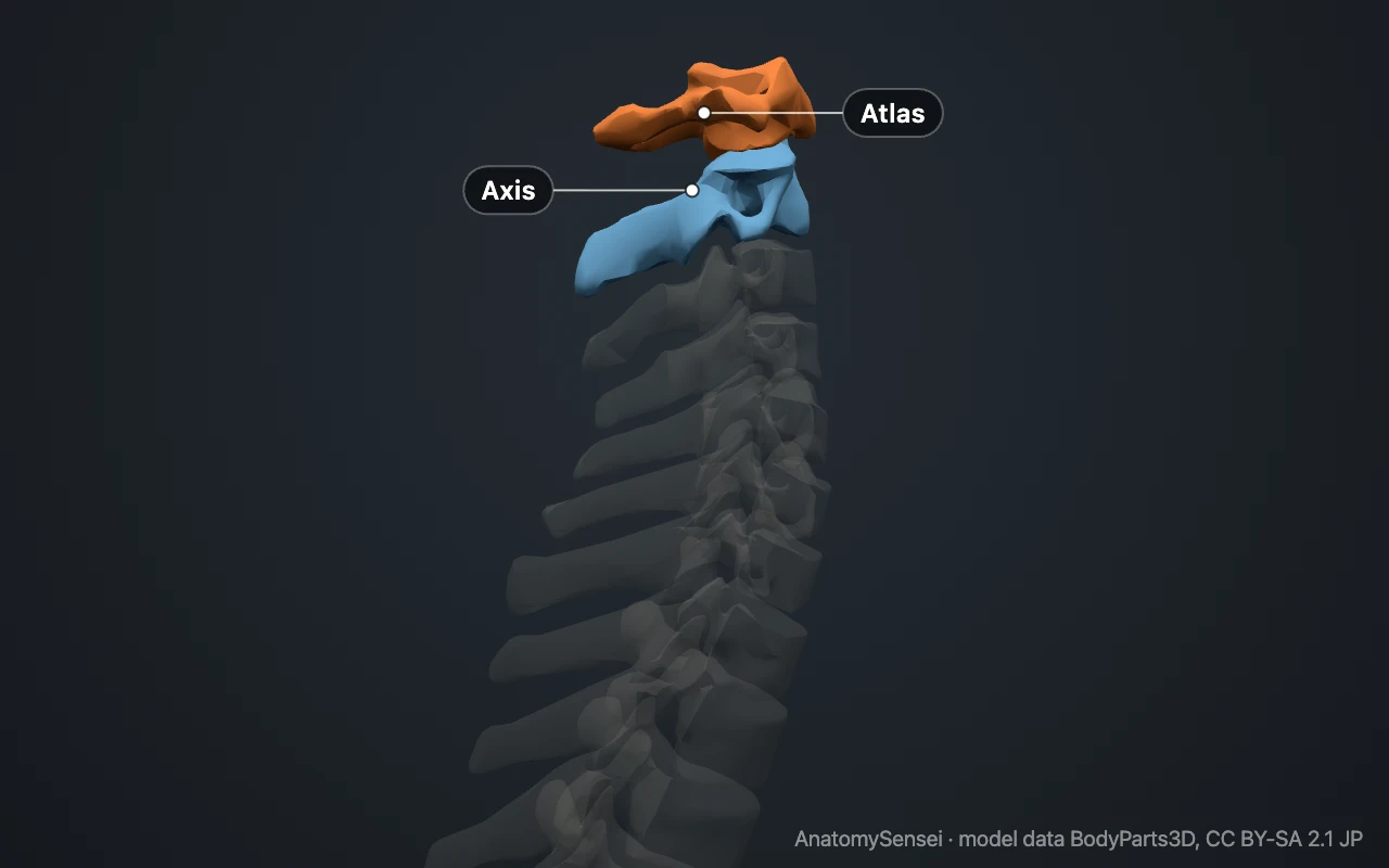
Below, the atlas meets the axis at three joints. Two lie between the lateral masses of the atlas and the upper facets of the axis. The third lies in the midline, where the back of the anterior arch touches the dens, the peg of the axis.
There is no disc above or below the atlas.
What attaches to the atlas?
The most important structure is the transverse ligament of the atlas. It stretches between the two lateral masses behind the dens and holds the peg against the anterior arch. It divides the ring into a small front part for the dens and a larger back part for the spinal cord.
Membranes connect the arches of the atlas to the rim of the opening in the skull above. Several small muscles that make fine adjustments to head position run from the atlas to the skull and to the axis.
What does the atlas do?
The atlas carries the head. Its joints with the skull allow nodding, the "yes" movement, and a little tilting to each side.
When the head turns, the atlas and the skull move together as one unit and pivot around the dens of the axis. This is the "no" movement. The wide ring leaves room for the spinal cord to stay clear during these movements.
Common problems with the atlas
A heavy blow to the top of the head, as in diving into shallow water, can force the lateral masses apart and break the ring, typically through both arches. This burst fracture is called a Jefferson fracture.
If the transverse ligament is torn or weakened, the atlas can slide forward on the axis, a state called atlantoaxial instability. It can follow injury and also occurs in rheumatoid arthritis and Down syndrome. This page describes anatomy only.
Common questions
Why is the first vertebra called the atlas?
After Atlas of Greek myth, who carried the heavens on his shoulders. The bone carries the skull in the same way.
Does the atlas have a vertebral body?
No. During development, the part that would have become its body joins the axis instead and forms the dens.
Is there a disc between the atlas and the axis?
No. The first intervertebral disc lies between C2 and C3.
Which vertebra lets the head nod?
Nodding takes place mainly between the atlas and the skull. Turning the head takes place mainly between the atlas and the axis.
Is this 3D model of the atlas free?
Yes. The interactive 3D model on this page is free to view and use for study, with no account or download needed. Its geometry comes from the open BodyParts3D data set (CC BY-SA).
About this free 3D model of the atlas
The interactive 3D model on this page is free to use in your browser, with no sign-up. It is built from scan-based anatomy data, so every bone is a separate, correctly placed shape you can turn and inspect from any side.