
Cranial bones
Ossa cranii
The cranial bones are the eight bones that enclose the brain. They are the frontal bone, two parietal bones, the occipital bone, two temporal bones, the sphenoid and the ethmoid.
Loading 3D model…
Free 3D model of the cranial bones. Whole skull. Drag to rotate, zoom with + and − (or Ctrl + scroll), click a bone to highlight and name it.
Key points
- Eight cranial bones form the braincase, or neurocranium.
- Four are single (frontal, occipital, sphenoid, ethmoid) and two are paired (parietal, temporal).
- They are joined by sutures, which are immovable joints in the adult.
- The sphenoid and ethmoid lie deep in the skull base and are mostly hidden from outside.
What are the cranial bones?
The cranial bones are the eight bones of the braincase. Together they form a closed box with a domed roof and an uneven floor. This box is called the neurocranium.
The roof, or calvaria, is made of flat, curved plates. The floor, or cranial base, is thicker and more complex, because nerves and blood vessels pass through it and the face and neck attach to it.
What are the names of the 8 cranial bones?
The frontal bone forms the forehead and the roofs of the eye sockets. Behind it, the two parietal bones form most of the roof and upper sides of the skull.
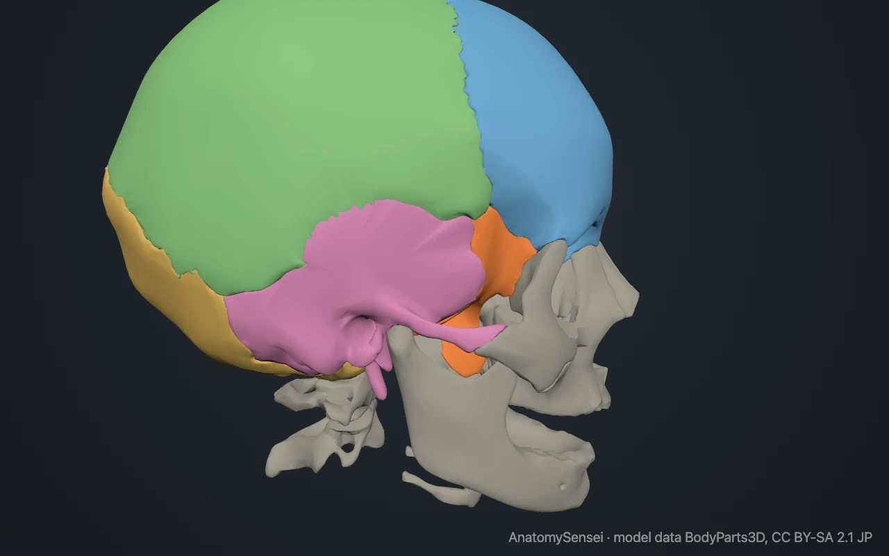
The occipital bone forms the back and part of the base. It contains the large opening through which the brain connects with the spinal cord. The two temporal bones form the lower sides and house the organs of hearing and balance.
The sphenoid is a butterfly-shaped bone that spans the base of the skull from side to side. The ethmoid is a light, delicate bone between the eye sockets, at the roof of the nose.
Where are the cranial bones?
They make up the upper and back part of the head. Everything that can be felt under the scalp belongs to a cranial bone, as does the forehead.
Several can be found by touch. Run a hand from the eyebrows up over the forehead for the frontal bone, then over the crown for the parietal bones. The firm bump at the back of the head above the neck is on the occipital bone, and the hard lump behind each ear is the mastoid process of the temporal bone.
Why are the sphenoid and ethmoid hard to see?
Both lie in the middle of the skull base, covered by the bones of the face in front and the other cranial bones at the sides. From outside, only a small part of the sphenoid shows, in the temple and at the back of the eye socket.
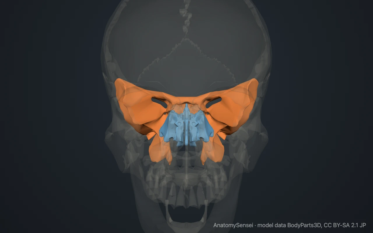
The sphenoid touches every other cranial bone. This is why it is often called the keystone of the skull. The ethmoid sits just in front of it and separates the brain from the nasal cavity.
What are the main sutures between the cranial bones?
A suture is a fibrous joint between two skull bones. Four are large enough to learn by name.
The coronal suture joins the frontal bone to the two parietal bones. The sagittal suture joins the two parietal bones along the midline. The lambdoid suture joins the parietal bones to the occipital bone, and the squamous suture on each side joins a parietal bone to a temporal bone.
On the side of the skull, the frontal, parietal, temporal and sphenoid bones meet at a small H-shaped area called the pterion. The bone is thin here.
How do the cranial bones differ in a baby?
At birth the bones of the skullcap are separated by wide, flexible sutures. Where several bones meet there are larger membrane-covered gaps called fontanelles, or soft spots.
The largest is the anterior fontanelle, between the frontal and parietal bones. The gaps let the skull bones overlap slightly during birth and give the brain room to grow. They close in early childhood. The model shows an adult skull.
What do the cranial bones do?
Their main job is to protect the brain. The curved plates of the skullcap spread the force of a blow over a wide area.
They also protect the inner ear, form the upper parts of the eye sockets and nasal cavity, and give attachment to the muscles of the scalp, jaw and neck. Openings in the cranial base let the spinal cord, cranial nerves and blood vessels pass in and out.
Common problems with the cranial bones
Skull fractures may be simple cracks in the skullcap, depressed fractures in which a piece is pushed inward, or fractures of the cranial base. A fracture at the pterion can tear an artery that runs just inside the bone.
In craniosynostosis, one or more sutures close too early in infancy, which changes the shape of the growing head. This page describes anatomy only.
Common questions
How many cranial bones are there?
Eight: one frontal, two parietal, one occipital, two temporal, one sphenoid and one ethmoid.
What is the difference between cranial and facial bones?
Cranial bones surround the brain. The 14 facial bones form the face and jaws and do not enclose the brain.
Which cranial bones are paired?
The parietal and temporal bones. There is one of each on the left and one on the right.
Which cranial bone touches all the others?
The sphenoid. It articulates with the frontal, both parietal, both temporal, the occipital and the ethmoid bones.
Is this 3D model of the cranial bones free?
Yes. The interactive 3D model on this page is free to view and use for study, with no account or download needed. Its geometry comes from the open BodyParts3D data set (CC BY-SA).
About this free 3D model of the cranial bones
The interactive 3D model on this page is free to use in your browser, with no sign-up. It is built from scan-based anatomy data, so every bone is a separate, correctly placed shape you can turn and inspect from any side.