
Frontal bone (forehead bone)
Os frontale
The frontal bone is the bone of the forehead. It forms the front of the braincase and the roof of each eye socket, and it contains the frontal sinuses.
Loading 3D model…
Free 3D model of the frontal bone. Whole skull. Drag to rotate, zoom with + and − (or Ctrl + scroll), click a bone to highlight and name it.
Key points
- The frontal bone forms the forehead and the upper rim and roof of the eye sockets.
- It is a single bone that develops from two halves.
- It meets the two parietal bones at the coronal suture.
- Two air spaces, the frontal sinuses, lie inside it above the nose.
What is the frontal bone?
The frontal bone is the single cranial bone at the front of the skull. Its shell-shaped upper part forms the forehead, and two thin plates at its lower edge form the roofs of the eye sockets.
It protects the frontal lobes of the brain, which rest directly behind and above it.
Where is the frontal bone?
It lies above the eyes and nose and in front of the crown. It reaches from the eyebrows up to a line that crosses the top of the head roughly from ear to ear.
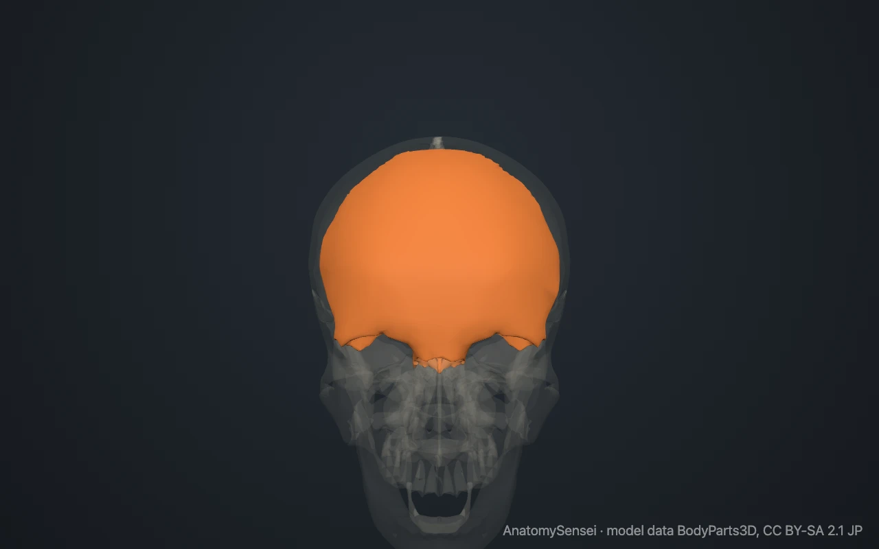
It is easy to feel. The whole forehead is frontal bone. Press a fingertip along the bony ridge under the eyebrow to trace the upper rim of the eye socket, which is the supraorbital margin.
What are the parts of the frontal bone?
The large curved plate of the forehead is the squamous part. The two horizontal plates that roof the eye sockets are the orbital parts. A small nasal part lies between them and meets the bones of the nose.
The supraorbital margin is the curved upper rim of each eye socket. A small notch or hole in it carries a nerve and vessels to the forehead. Above each margin is a low ridge under the eyebrow, the superciliary arch, and the smooth area between the two ridges is the glabella.
At its outer end each margin runs down into a short zygomatic process, which meets the cheekbone. These landmarks are part of the one bone and are not separate pieces in the model.
What does the frontal bone articulate with?
It meets twelve bones. Behind, it joins the two parietal bones along the coronal suture. Below and behind, it joins the sphenoid and the ethmoid.
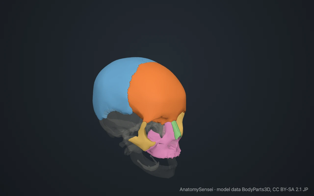
In the face it joins the two nasal bones, the two maxillae, the two lacrimal bones and the two zygomatic bones. Its joints with the nasal bones and maxillae form the bridge of the nose.
What are the frontal sinuses?
The frontal sinuses are two air-filled spaces inside the frontal bone, above the nose and behind the inner ends of the eyebrows. Each drains into the nasal cavity.
They are absent at birth and grow during childhood. Their size and shape differ from person to person, and the left and right are rarely the same.
What attaches to the frontal bone?
Part of the temporalis muscle, a chewing muscle, arises from the side of the frontal bone in the temple. Small muscles of the eyebrow and eyelid attach near the inner end of the supraorbital margin.
Inside the skull, a ridge in the midline called the frontal crest anchors the falx cerebri. This is the fold of tough membrane that separates the two halves of the brain.
Is the frontal bone one bone or two?
In an adult it is one bone. It begins as a left and a right half, separated by a suture in the midline of the forehead called the frontal or metopic suture.
The two halves normally fuse in early childhood. In some adults the suture remains visible, which is a harmless variant.
Common problems with the frontal bone
The forehead is thick and strong, so frontal bone fractures usually follow a hard impact. They may involve the wall of a frontal sinus or the roof of an eye socket.
Frontal sinusitis is inflammation of the lining of the frontal sinuses and causes pain above the eyes. This page describes anatomy only.
Common questions
Is the forehead a single bone?
Yes. In adults the forehead is the frontal bone. It forms from two halves that fuse in early childhood.
What is the glabella?
The smooth, slightly raised area of the frontal bone between the eyebrows, just above the nose.
Which suture is behind the frontal bone?
The coronal suture, where the frontal bone meets the two parietal bones.
Does the frontal bone form part of the eye socket?
Yes. It forms the roof and the upper rim of each eye socket.
Is this 3D model of the frontal bone free?
Yes. The interactive 3D model on this page is free to view and use for study, with no account or download needed. Its geometry comes from the open BodyParts3D data set (CC BY-SA).
About this free 3D model of the frontal bone
The interactive 3D model on this page is free to use in your browser, with no sign-up. It is built from scan-based anatomy data, so every bone is a separate, correctly placed shape you can turn and inspect from any side.