
Ethmoid bone
Os ethmoideum
The ethmoid is a small, light bone between the eye sockets, at the roof of the nose. It separates the nasal cavity from the brain, and the nerves of smell pass through its sieve-like upper plate.
Loading 3D model…
Free 3D model of the ethmoid bone. Whole skull. Drag to rotate, zoom with + and − (or Ctrl + scroll), click a bone to highlight and name it.
Key points
- The ethmoid lies deep in the face, between the two eye sockets.
- Its cribriform plate is pierced by small holes for the nerves of smell.
- It forms part of the nasal septum, the inner wall of each eye socket and the roof of the nose.
- It is full of small air spaces called ethmoidal air cells.
What is the ethmoid bone?
The ethmoid is a single cranial bone in the midline of the face. Its name comes from the Greek for sieve, after the many small holes in its upper plate.
It is roughly cube-shaped and very light, because most of it is thin plates of bone around air-filled spaces.
Where is the ethmoid bone?
It lies between the eye sockets, behind the bridge of the nose and below the frontal bone. The sphenoid is directly behind it.
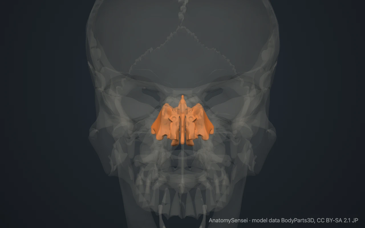
It cannot be felt from outside. The nearest point on the surface is the inner corner of each eye, where the ethmoid lies a short distance behind the lacrimal bone. The model on this page opens with the other bones see-through so that the ethmoid can be seen.
What are the parts of the ethmoid?
The cribriform plate is a horizontal strip that forms the roof of the nasal cavity and a small part of the floor of the braincase. A fin of bone, the crista galli, stands up from its midline.
The perpendicular plate hangs down from the cribriform plate in the midline. It forms the upper part of the nasal septum, the wall between the left and right sides of the nose.
On each side hangs an ethmoidal labyrinth, a block of thin-walled air cells. Its smooth outer face forms part of the inner wall of the eye socket. Its inner face carries two curled shelves, the superior and middle nasal conchae, which project into the nasal cavity.
What does the ethmoid articulate with?
It meets two cranial bones. The frontal bone lies above and in front of it, and the sphenoid lies behind.
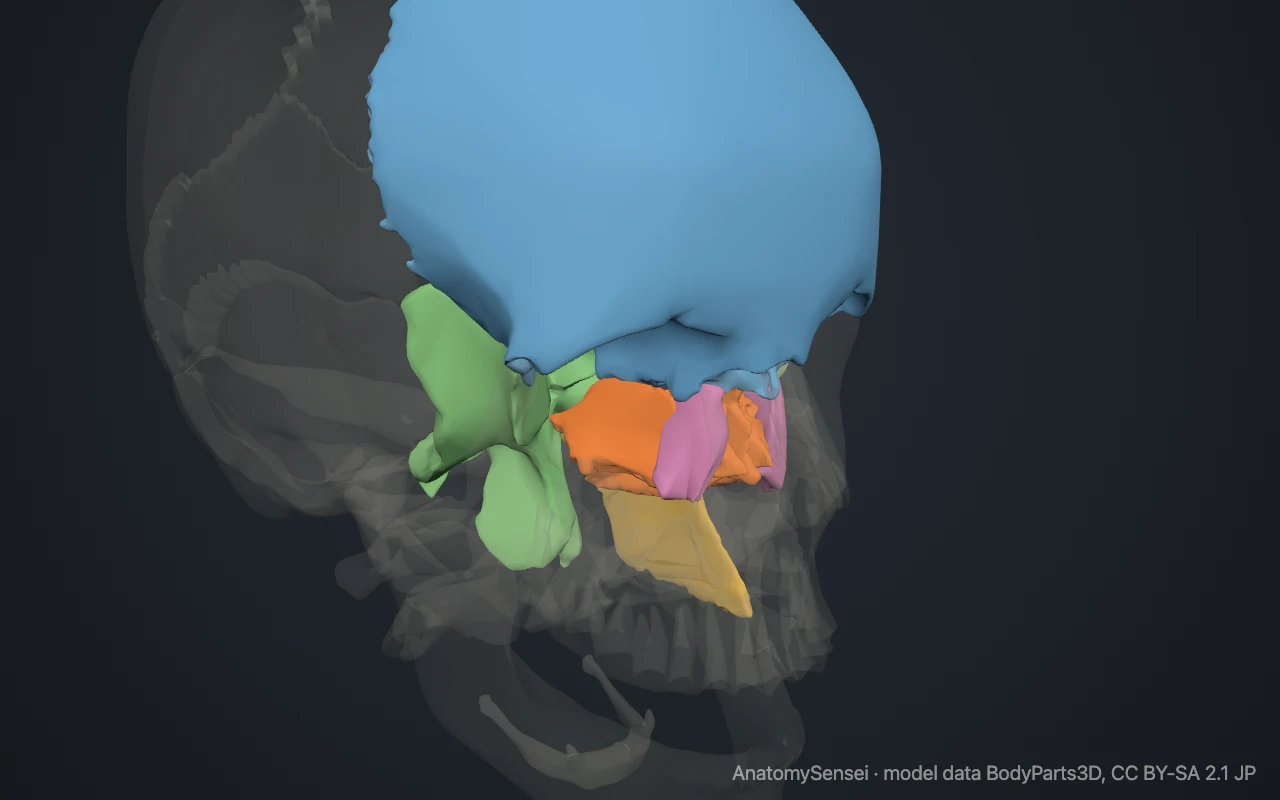
It also meets eleven facial bones: the two nasal bones, the two maxillae, the two lacrimal bones, the two palatine bones, the two inferior nasal conchae and the vomer. The vomer continues the nasal septum downward from the perpendicular plate.
What are the ethmoidal air cells?
The ethmoidal air cells are a honeycomb of small air spaces inside each labyrinth. Together they are called the ethmoidal sinuses, one of the four pairs of sinuses around the nose.
They open into the nasal cavity. Their number and size vary from person to person.
What does the ethmoid do?
It separates the nasal cavity from the brain and from the eye sockets. The fine nerves of smell pass up from the lining of the nose through the holes of the cribriform plate to reach the brain.
Its conchae increase the surface area inside the nose, which helps to warm and moisten air as it is breathed in. The crista galli anchors the front end of the falx cerebri, the membrane between the two halves of the brain.
Common problems with the ethmoid
The ethmoid is thin and fragile. A hard blow to the nose or forehead can fracture the cribriform plate, which may damage the nerves of smell or let the fluid that surrounds the brain leak into the nose.
The paper-thin plate in the inner wall of the eye socket can break in injuries to the eye region. Ethmoid sinusitis is inflammation of the lining of the air cells and causes pain between the eyes. This page describes anatomy only.
Common questions
Is the ethmoid a cranial or a facial bone?
It is counted as a cranial bone, because its cribriform plate forms part of the floor of the braincase.
Why is it called the ethmoid?
From the Greek for sieve. The cribriform plate is full of small holes.
What is the crista galli?
A small upright fin of bone on top of the ethmoid, in the midline. The name means the crest of a cock.
Are the nasal conchae part of the ethmoid?
The superior and middle conchae are. The inferior nasal concha on each side is a separate facial bone.
Is this 3D model of the ethmoid bone free?
Yes. The interactive 3D model on this page is free to view and use for study, with no account or download needed. Its geometry comes from the open BodyParts3D data set (CC BY-SA).
About this free 3D model of the ethmoid bone
The interactive 3D model on this page is free to use in your browser, with no sign-up. It is built from scan-based anatomy data, so every bone is a separate, correctly placed shape you can turn and inspect from any side.