
Lacrimal bone
Os lacrimale
The lacrimal bone is a thin plate of bone about the size of a fingernail in the inner wall of each eye socket. It is the smallest of the facial bones and helps to form the channel that drains tears into the nose.
Loading 3D model…
Free 3D model of the lacrimal bone. Whole skull. Drag to rotate, zoom with + and − (or Ctrl + scroll), click a bone to highlight and name it.
- Maxilla
Key points
- The lacrimal bone is the smallest and most fragile bone of the face.
- It sits at the front of the inner wall of the eye socket.
- It joins the frontal bone, ethmoid, maxilla and inferior nasal concha.
- With the maxilla it forms the hollow for the tear sac and the canal that carries tears to the nose.
What is the lacrimal bone?
The lacrimal bone is a small, thin, roughly rectangular facial bone. There is one in each eye socket. It is the smallest of the 14 facial bones and one of the most delicate bones in the body.
Its name comes from the Latin word for a tear. The bone lies beside the tear sac and forms part of the path that tears take from the eye to the nose.
Where is the lacrimal bone?
It lies at the front of the inner wall of the orbit, the wall nearest the nose. It sits just behind the frontal process of the maxilla, which forms the inner rim of the eye socket.
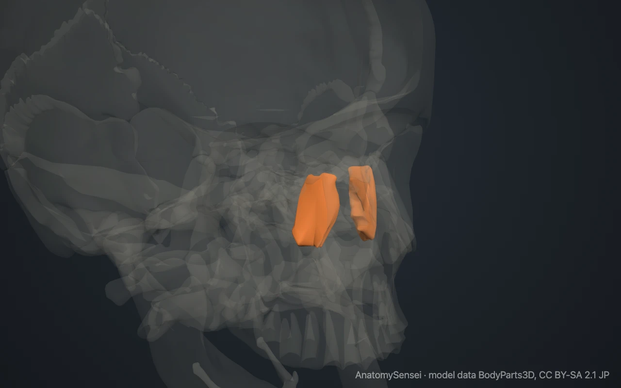
The lacrimal bone cannot be felt through the skin, because it lies inside the socket behind the rim. Its position is roughly level with the inner corner of the eye, a short distance back. In the model, the rest of the skull is made see-through so that the bone shows.
What are the parts of the lacrimal bone?
The bone has two surfaces and four borders. The outer surface faces into the orbit and is divided by a vertical ridge called the posterior lacrimal crest.
In front of the crest is a vertical groove, the lacrimal groove. Behind the crest the surface is flat and forms part of the smooth inner wall of the orbit. The lower end of the crest curves forward into a small hook called the lacrimal hamulus.
The inner surface faces the nose. It forms a small part of the side wall of the nasal cavity and covers some of the front air cells of the ethmoid bone.
What does the lacrimal bone articulate with?
Each lacrimal bone joins four bones. Above, it meets the frontal bone. Behind, it meets the thin orbital plate of the ethmoid bone. In front and below, it meets the maxilla.
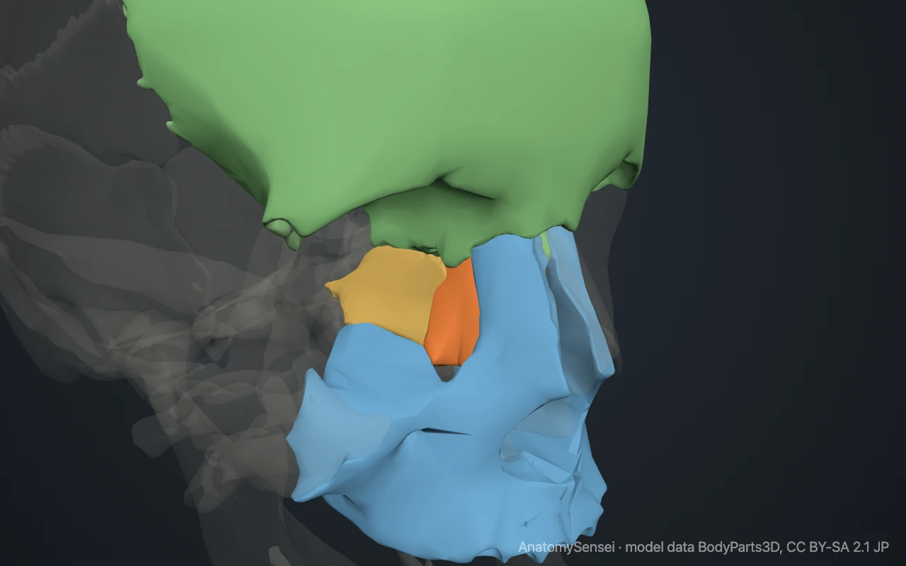
The fourth is the inferior nasal concha. A small downward process of the lacrimal bone reaches into the nose to meet it, and the two help to wall in the tear canal. All of these joints are fixed sutures.
What does the lacrimal bone form part of?
It forms part of three things. It is one of the seven bones of the orbit, where it completes the front of the inner wall. It forms a small part of the side wall of the nasal cavity.
It also forms part of the tear drainage pathway. The lacrimal groove joins a matching groove on the maxilla to make a hollow called the fossa for the lacrimal sac. Below this the two bones, with the inferior nasal concha, enclose the nasolacrimal canal.
What passes through and attaches to it?
Tears collect at the inner corner of the eye and drain into the lacrimal sac, which rests in the hollow formed by the lacrimal bone and the maxilla. From the sac, the nasolacrimal duct runs down through the nasolacrimal canal and opens into the nose below the inferior nasal concha. This is why the nose runs during crying.
A small deep part of the orbicularis oculi, the muscle that closes the eyelids, attaches to the posterior lacrimal crest. Its pull on the tear sac during blinking helps to move tears along.
Common problems with the lacrimal bone
The lacrimal bone is very thin, so it can break in fractures of the inner wall of the orbit and in injuries that crush the area between the eyes, known as naso-orbito-ethmoid fractures.
Damage or narrowing along the bony canal can lead to a blocked tear duct, in which tears spill over the eyelid instead of draining. This page describes anatomy only.
Common questions
Is the lacrimal bone the smallest bone of the face?
Yes. It is the smallest of the 14 facial bones. The smallest bones in the whole body are the ossicles of the ear.
Does the lacrimal bone make tears?
No. Tears are made by the lacrimal gland, which lies in the upper outer part of the orbit. The bone only helps to drain them.
How many lacrimal bones are there?
Two, one in the inner wall of each eye socket.
Can the lacrimal bone be felt?
No. It lies inside the eye socket behind the bony rim.
Is this 3D model of the lacrimal bone free?
Yes. The interactive 3D model on this page is free to view and use for study, with no account or download needed. Its geometry comes from the open BodyParts3D data set (CC BY-SA).
About this free 3D model of the lacrimal bone
The interactive 3D model on this page is free to use in your browser, with no sign-up. It is built from scan-based anatomy data, so every bone is a separate, correctly placed shape you can turn and inspect from any side.