
Maxilla (upper jaw bone)
Maxilla
The maxilla is the upper jaw bone. There are two, a left and a right, joined in the midline below the nose. Together they carry the upper teeth and form the centre of the face.
Loading 3D model…
Free 3D model of the maxilla. Whole skull. Drag to rotate, zoom with + and − (or Ctrl + scroll), click a bone to highlight and name it.
Key points
- The two maxillae form the upper jaw and carry the upper teeth.
- Each maxilla has a hollow body and four processes: frontal, zygomatic, alveolar and palatine.
- It forms part of the floor of the orbit, the side wall and floor of the nose, and the front of the hard palate.
- The maxilla touches every other facial bone except the mandible.
What is the maxilla?
The maxilla is the bone of the upper jaw. It is a paired bone, and the left and right maxillae meet at a seam in the midline called the intermaxillary suture. The plural is maxillae.
After the mandible, it is the largest bone of the face. It is also the most central. Each maxilla reaches up to the eye socket, inward to the nose and down to the mouth, so it helps to form all three.
Where is the maxilla?
It lies in the middle of the face, between the eye sockets above and the mouth below, on each side of the nose.
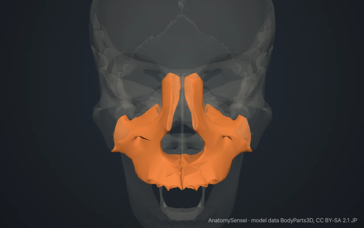
You can feel it on your own face. Press on the skin between the upper lip and the nose, then move the finger sideways below the cheekbone. The firm bone under the finger is the maxilla. Inside the mouth, the gum around the upper teeth and the front of the roof of the mouth also cover the maxilla.
What are the parts of the maxilla?
Each maxilla has a body and four processes. The body is shaped roughly like a pyramid and is hollow. The space inside it is the maxillary sinus, the largest of the air sinuses around the nose.
The frontal process runs upward beside the nose to reach the frontal bone. The zygomatic process points outward to meet the cheekbone. The alveolar process is the curved ridge that carries the upper teeth, which the model does not include. The palatine process is a horizontal shelf that meets its partner in the midline to form the front of the hard palate.
Just below the rim of the eye socket is the infraorbital foramen, a small hole on the front of the body. In the midline at the bottom of the nasal opening, the two bones form a small forward point called the anterior nasal spine.
What does the maxilla articulate with?
Each maxilla joins nine bones. Two are cranial bones: the frontal bone and the ethmoid bone. Six are facial bones: the nasal, lacrimal and zygomatic bones, the palatine bone, the vomer and the inferior nasal concha. The ninth is the maxilla of the other side.
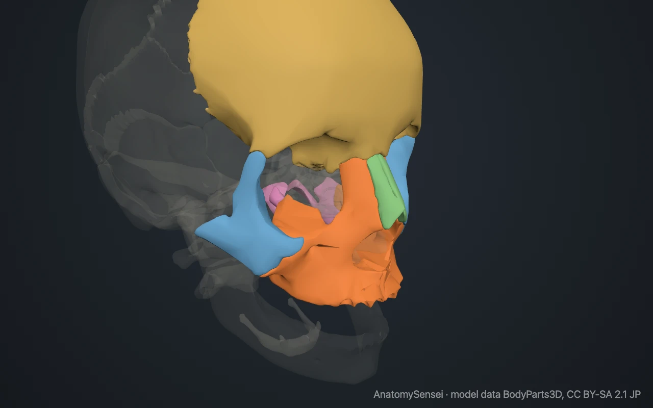
All of these joints are sutures and do not move. The upper teeth are held in sockets in the alveolar process. The maxilla has no direct joint with the mandible. The two jaws meet only through the teeth.
What does the maxilla form part of?
The upper surface of the body forms most of the floor of the orbit. The inner surface forms much of the side wall of the nasal cavity, and has the opening of the maxillary sinus.
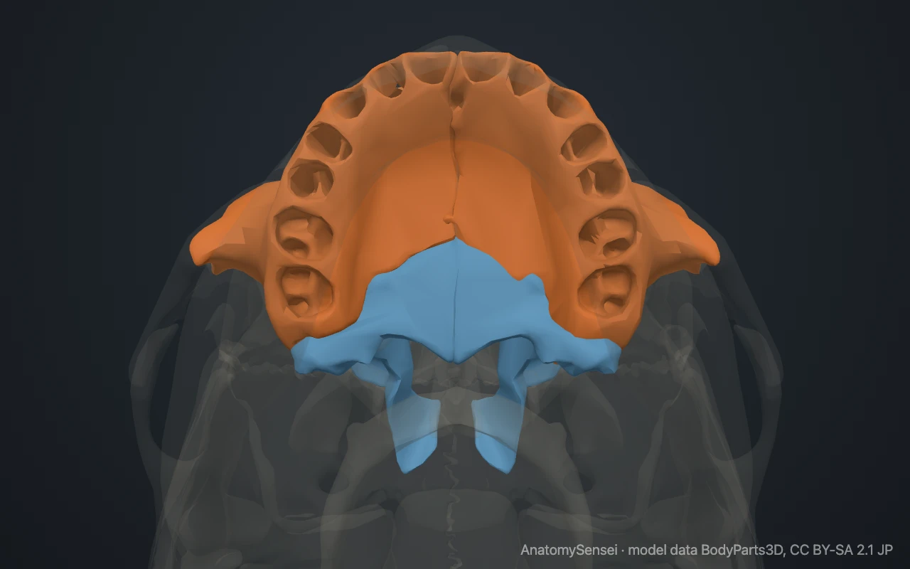
The palatine processes form the front and larger part of the hard palate, which is the roof of the mouth and also the floor of the nose. The back part of the hard palate comes from the palatine bones.
What passes through and attaches to the maxilla?
The infraorbital nerve and its vessels run forward in a groove and canal in the floor of the orbit and come out at the infraorbital foramen. This nerve supplies feeling to the cheek, the side of the nose and the upper lip. Behind the upper front teeth, the incisive canals carry the nasopalatine nerves through the palate.
Several small muscles of facial expression arise from the front of the maxilla, including muscles that lift the upper lip. The buccinator, the muscle of the cheek, arises partly from the alveolar process, and part of the masseter arises from the zygomatic process.
Common problems with the maxilla
Fractures across the middle of the face that involve the maxillae are grouped into three patterns named Le Fort fractures. The thin floor of the orbit can also break on its own, which is called a blow-out fracture.
When the palatine processes fail to join before birth, the result is a cleft palate. The roots of the upper back teeth lie close to the floor of the maxillary sinus, so problems with these teeth and with the sinus can affect each other. This page describes anatomy only.
Common questions
Is the maxilla one bone or two?
Two. A left and a right maxilla join in the midline to form the upper jaw.
Does the maxilla move?
No. It is fixed to the rest of the skull. Only the lower jaw, the mandible, moves during chewing.
What is the maxillary sinus?
An air-filled space inside the body of the maxilla, below the eye and beside the nose. It opens into the nasal cavity.
What is the difference between the maxilla and the mandible?
The maxilla is the fixed upper jaw. The mandible is the movable lower jaw.
Is this 3D model of the maxilla free?
Yes. The interactive 3D model on this page is free to view and use for study, with no account or download needed. Its geometry comes from the open BodyParts3D data set (CC BY-SA).
About this free 3D model of the maxilla
The interactive 3D model on this page is free to use in your browser, with no sign-up. It is built from scan-based anatomy data, so every bone is a separate, correctly placed shape you can turn and inspect from any side.