
Mandible (lower jaw bone)
Mandibula
The mandible is the lower jaw bone. It is the largest and strongest bone of the face and the only one that moves, hinging on the temporal bones just in front of the ears.
Loading 3D model…
Free 3D model of the mandible. Whole skull. Drag to rotate, zoom with + and − (or Ctrl + scroll), click a bone to highlight and name it.
Key points
- The mandible is the lower jaw and carries the lower teeth.
- It has a horseshoe-shaped body and an upright ramus on each side.
- It joins only the two temporal bones, at the temporomandibular joints.
- All four chewing muscles attach to the mandible.
What is the mandible?
The mandible is the bone of the lower jaw. It is a single bone that forms the chin and the jawline and carries the lower teeth. The model shows the bone without teeth.
It begins as two halves, left and right, which fuse at the chin in early childhood. It is the only bone of the face that moves, and its movement is what opens and closes the mouth.
Where is the mandible?
It lies at the bottom of the face, below the upper jaw. Its two ends reach up to the base of the skull just in front of each ear.
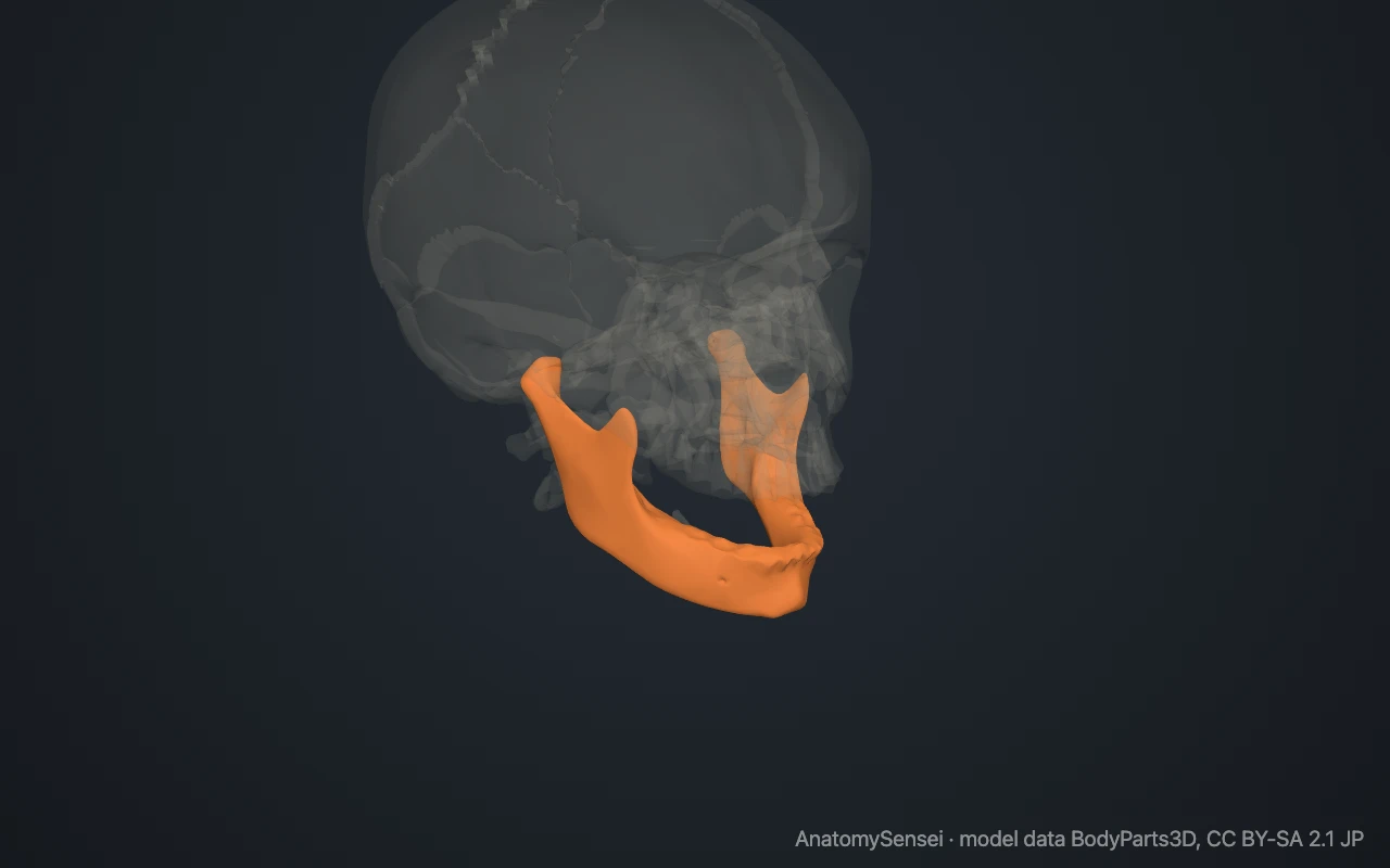
The whole lower border can be felt on your own face. Start at the point of the chin and follow the edge of the jaw backward to the corner below the ear, which is the angle of the mandible. Then place a fingertip just in front of the ear opening and open the mouth. The knob that slides forward under the finger is the head of the mandible.
What are the parts of the mandible?
The mandible has a horizontal body and two upright plates, each called a ramus. The body is curved like a horseshoe. Its upper edge, the alveolar part, holds the sockets for the lower teeth. The body and ramus meet at the angle of the mandible.
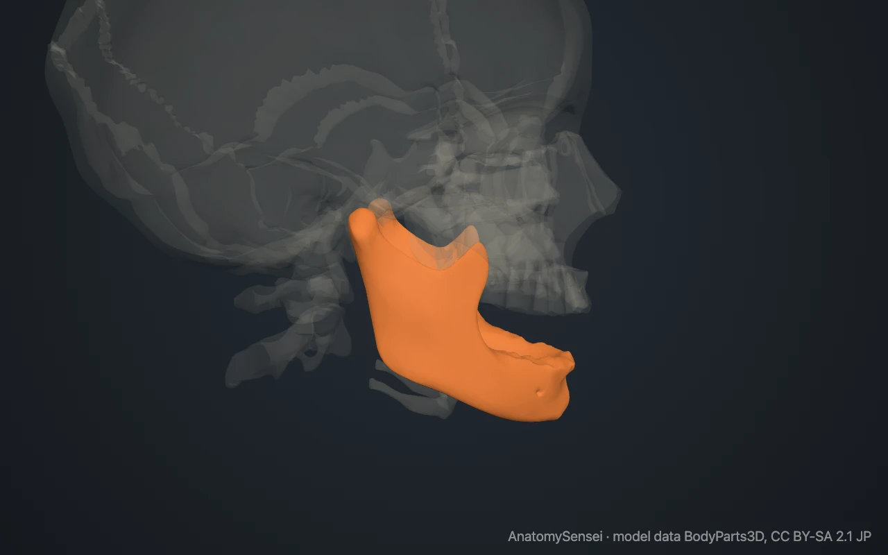
The top of each ramus ends in two processes separated by a dip called the mandibular notch. The front one is the coronoid process, a thin triangle of bone. The back one is the condylar process, which has a narrow neck and a rounded head.
The front of the body has a raised area at the chin called the mental protuberance. On each side of it, below the premolar teeth, is the mental foramen. On the inner side of each ramus is the mandibular foramen, the entrance to a canal that runs through the bone.
What does the mandible articulate with?
The mandible joins only the two temporal bones. On each side, the head of the mandible fits into a hollow on the underside of the temporal bone called the mandibular fossa. This is the temporomandibular joint, often shortened to TMJ.
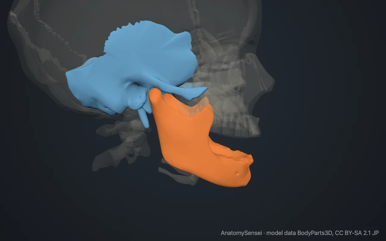
The joint is a synovial joint with a small disc of cartilage between the two bones. It lets the jaw hinge open and also slide forward and from side to side. The two joints always work together, because both are on the same bone.
What attaches to the mandible?
All four muscles of mastication, the chewing muscles, attach to it. The masseter attaches to the outer surface of the ramus and angle. The temporalis attaches to the coronoid process. The medial pterygoid attaches to the inner surface of the angle, and the lateral pterygoid attaches to the neck of the condylar process.
Muscles of the floor of the mouth and the tongue attach to the inner surface of the body. These include the mylohyoid, the geniohyoid, the genioglossus and the front belly of the digastric. Several muscles of the lower lip and chin attach to the outer surface.
What passes through the mandible?
The inferior alveolar nerve and its vessels enter the bone at the mandibular foramen and run forward inside the mandibular canal, below the roots of the teeth. The nerve supplies feeling to the lower teeth.
Its end branch, the mental nerve, leaves through the mental foramen to supply the skin of the chin and the lower lip.
What does the mandible do?
It moves the lower teeth against the upper teeth for biting and chewing. It also takes part in speech and forms the bony frame for the floor of the mouth and the tongue.
Common problems with the mandible
The mandible is one of the facial bones broken most often. Fractures commonly occur at the neck of the condylar process, at the angle and in the body near the chin. Because the bone is a ring-like arch, it often breaks in two places at once.
In a dislocated jaw, the head of the mandible slips forward out of the mandibular fossa and the mouth cannot close. Pain or clicking at the joint is grouped under the name temporomandibular disorders. This page describes anatomy only.
Common questions
Is the mandible the strongest bone of the face?
Yes. It is the largest and strongest facial bone.
Is the mandible one bone or two?
One in adults. It starts as a left and a right half that fuse at the chin in early childhood.
Which bone does the mandible join?
The temporal bone on each side, at the temporomandibular joint in front of the ear.
What is the angle of the mandible?
The corner below the ear where the body of the mandible turns upward into the ramus.
Is this 3D model of the mandible free?
Yes. The interactive 3D model on this page is free to view and use for study, with no account or download needed. Its geometry comes from the open BodyParts3D data set (CC BY-SA).
About this free 3D model of the mandible
The interactive 3D model on this page is free to use in your browser, with no sign-up. It is built from scan-based anatomy data, so every bone is a separate, correctly placed shape you can turn and inspect from any side.