
Temporal bone
Os temporale
The temporal bones are a pair of bones at the lower sides of the skull, around the ears. Each contains the organs of hearing and balance and forms the socket of the jaw joint.
Loading 3D model…
Free 3D model of the temporal bone. Whole skull. Drag to rotate, zoom with + and − (or Ctrl + scroll), click a bone to highlight and name it.
Key points
- There are two temporal bones, one around each ear.
- Each houses the middle and inner ear inside its dense petrous part.
- The mandible hinges on the temporal bone at the temporomandibular joint.
- The lump behind the ear is the mastoid process of the temporal bone.
What is the temporal bone?
The temporal bone is a paired cranial bone at the side and base of the skull. It is one of the most complex bones in the body, because it combines a thin protective plate, a joint surface, several muscle attachments and the chambers of the ear.
It protects the temporal lobe of the brain, which lies against its inner surface.
Where is the temporal bone?
It lies around and above the ear. The parietal bone is above it, the occipital bone behind it and the sphenoid in front. A wedge of the bone also runs inward across the floor of the skull.
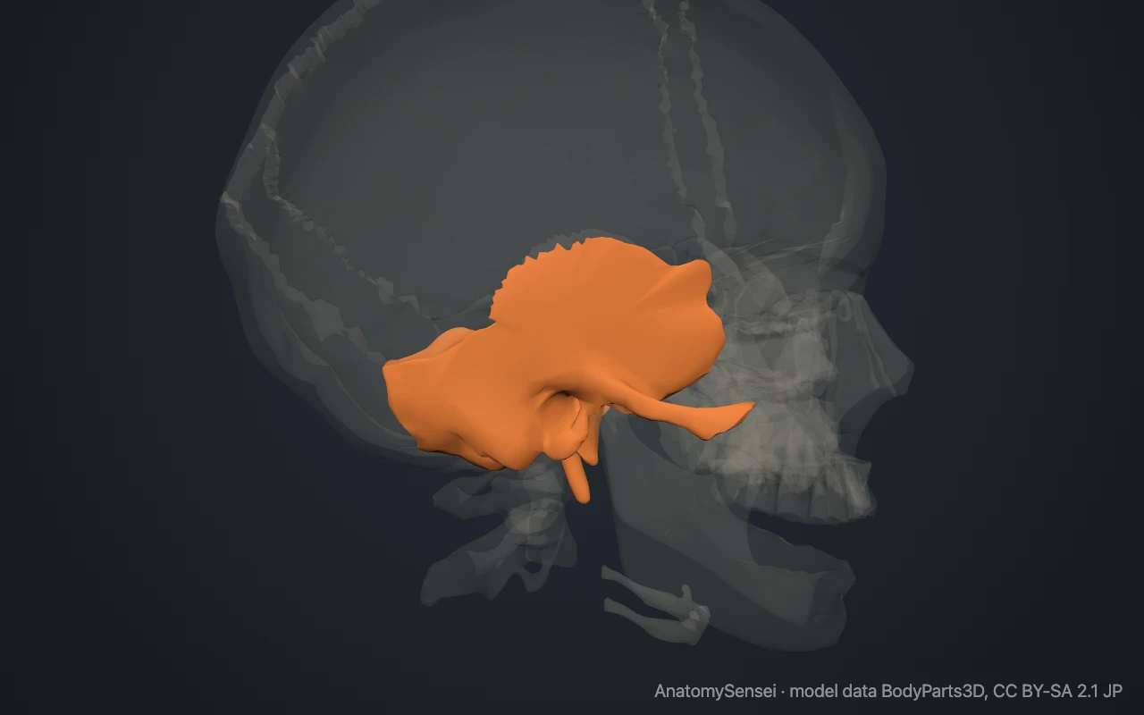
Several parts can be felt. The hard, rounded lump just behind the earlobe is the mastoid process. Place a fingertip just in front of the ear opening and open the mouth: the movement felt there is the head of the mandible sliding in its socket on the temporal bone.
What are the parts of the temporal bone?
The squamous part is the thin, fan-shaped plate above the ear. A slender bar, the zygomatic process, projects forward from it to meet the cheekbone. Under the root of this bar is the mandibular fossa, the hollow that receives the lower jaw.
The petrous part is a dense pyramid of bone that points inward and forward along the skull base. It encloses the middle and inner ear. Behind the ear it continues into the mastoid part, which ends below in the mastoid process and contains small air cells.
The tympanic part is a curved plate around the external acoustic meatus, the bony ear canal. Below it, the styloid process hangs down as a thin spike. These are regions of one bone and cannot be highlighted separately in the model.
What does the temporal bone articulate with?
Each temporal bone meets five bones. It joins the parietal bone above at the squamous suture, the occipital bone behind and the sphenoid in front.
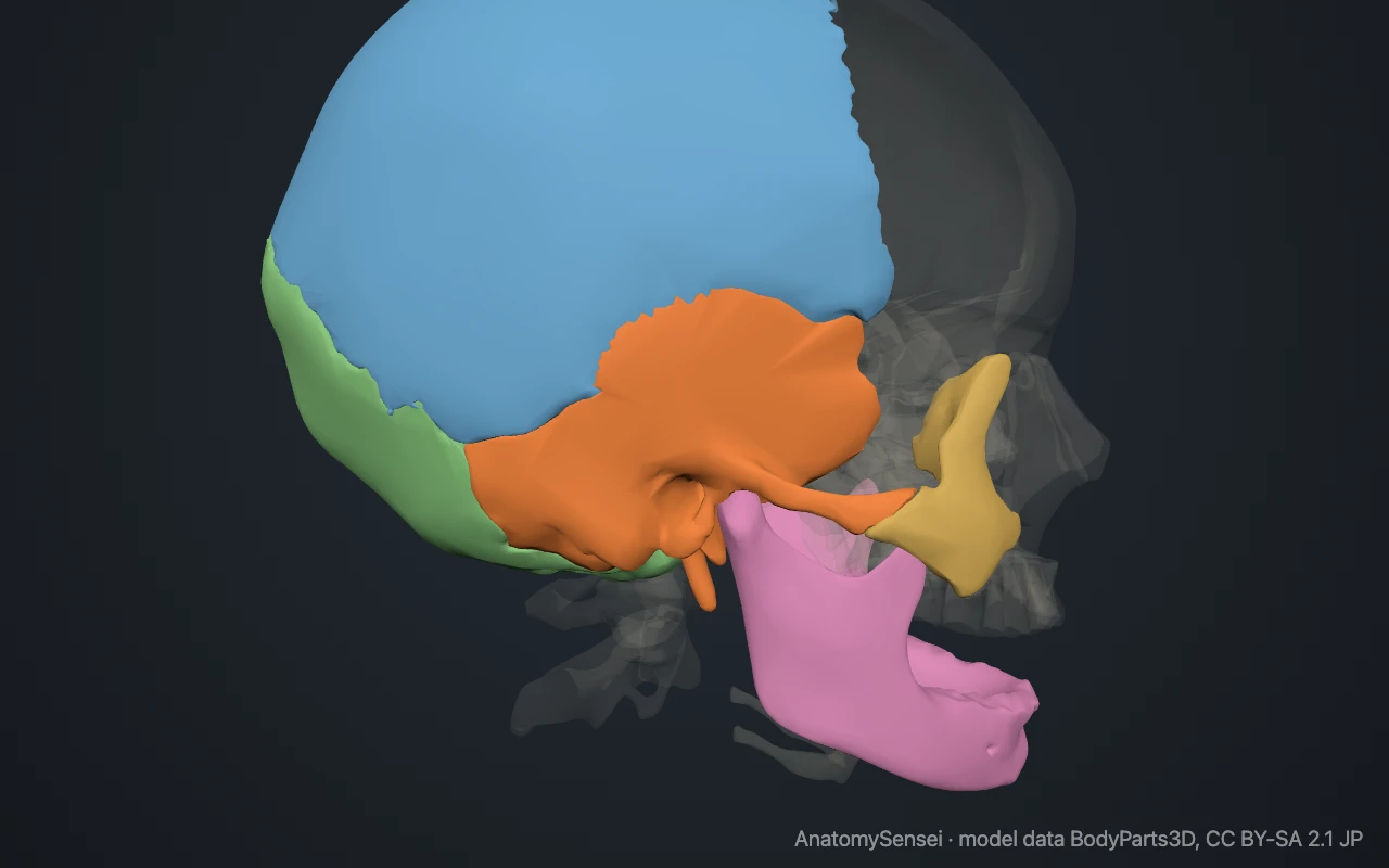
Its zygomatic process joins the zygomatic bone to form the zygomatic arch, the bony bar that can be felt running from the cheek to the ear.
The head of the mandible fits into the mandibular fossa to form the temporomandibular joint. This is the only freely movable joint between the skull bones shown in the model.
How is the temporal bone related to the ear?
The whole of the ear beyond the visible flap lies in this bone. The ear canal leads inward to the eardrum. Behind the eardrum is the middle ear, a small air-filled chamber crossed by three tiny bones that carry sound.
Deeper still, inside the petrous part, is the inner ear. It holds the cochlea for hearing and the semicircular canals for balance. The model shows the temporal bone from outside and does not include the three ear bones.
What attaches to the temporal bone?
The temporalis muscle covers the squamous part, and part of the masseter arises from the zygomatic arch. Both close the jaw.
The sternocleidomastoid, the large muscle at the side of the neck, attaches to the mastoid process. The styloid process anchors small muscles of the tongue and throat and the ligament from which the hyoid bone hangs.
Common problems with the temporal bone
Temporal bone fractures follow a hard blow to the side of the head and may damage hearing, balance or the facial nerve, which runs through a canal in the bone. The squamous part is thin and is more easily broken than the rest.
Mastoiditis is infection of the air cells in the mastoid part, usually spreading from the middle ear. Disorders of the temporomandibular joint cause pain and clicking in front of the ear. This page describes anatomy only.
Common questions
What is the bone behind the ear called?
The mastoid process. It is part of the temporal bone.
Is the temple the same as the temporal bone?
Not exactly. The temple is the region at the side of the forehead. Its bony floor is formed by the temporal bone together with parts of the frontal, parietal and sphenoid bones.
Which nerve runs through the temporal bone?
The facial nerve passes through it, and the nerve of hearing and balance ends inside it.
How many temporal bones are there?
Two, one on each side of the skull.
Is this 3D model of the temporal bone free?
Yes. The interactive 3D model on this page is free to view and use for study, with no account or download needed. Its geometry comes from the open BodyParts3D data set (CC BY-SA).
About this free 3D model of the temporal bone
The interactive 3D model on this page is free to use in your browser, with no sign-up. It is built from scan-based anatomy data, so every bone is a separate, correctly placed shape you can turn and inspect from any side.