
Parietal bone
Os parietale
The parietal bones are a pair of curved, four-sided plates that form most of the roof and upper sides of the skull. The left and right bones meet along the midline on top of the head.
Loading 3D model…
Free 3D model of the parietal bone. Whole skull. Drag to rotate, zoom with + and − (or Ctrl + scroll), click a bone to highlight and name it.
Key points
- There are two parietal bones, one on each side of the top of the head.
- Each is a four-sided plate with four borders and four corners.
- They meet each other at the sagittal suture.
- Each parietal bone joins five bones: its partner, the frontal, occipital, temporal and sphenoid.
What is the parietal bone?
The parietal bone is a paired flat bone of the skullcap. The name comes from the Latin for wall. The two bones together form the largest part of the roof of the skull.
Each is roughly square and gently domed, smooth and convex on the outside and hollow on the inside where it covers the brain.
Where are the parietal bones?
They lie on the top and upper sides of the head, behind the frontal bone and in front of the occipital bone. Below, each one meets the temporal bone above the ear.
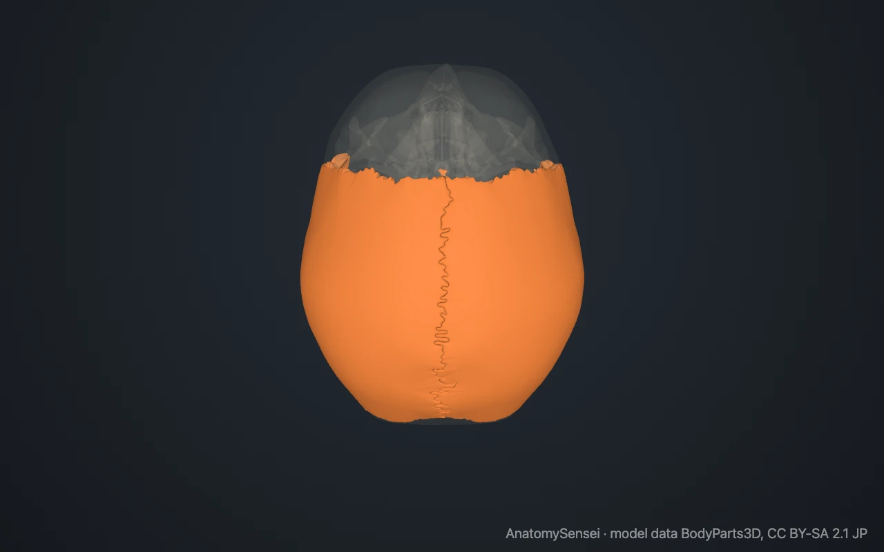
To find them, place both palms flat on the crown of the head. The bone under each palm is a parietal bone. The widest point of the head, a few finger-widths above and behind each ear, is the parietal eminence.
What are the landmarks of the parietal bone?
The outer surface carries the parietal eminence, the most bulging part of the bone. Below it, two faint curved lines cross the surface. These are the superior and inferior temporal lines, which mark the upper edge of the temple.
Near the back of the upper border there is often a small hole, the parietal foramen, for a small vein. The inner surface is marked by branching grooves made by the arteries that supply the covering of the brain.
The bone has four corners. The front upper corner lies at the bregma, where the coronal and sagittal sutures cross. The back upper corner lies at the lambda, where the sagittal and lambdoid sutures meet.
What does the parietal bone articulate with?
Each parietal bone meets five bones along four borders. Its upper border joins the other parietal bone at the sagittal suture. Its front border joins the frontal bone at the coronal suture.
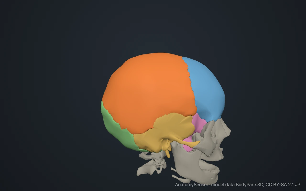
Its back border joins the occipital bone at the lambdoid suture. Its lower border joins the temporal bone at the squamous suture, where the thin edge of the temporal bone overlaps the parietal.
The front lower corner touches the greater wing of the sphenoid. This meeting point in the temple is part of the pterion.
What attaches to the parietal bone?
The temporalis muscle, which closes the jaw, arises from the side of the bone below the inferior temporal line. The sheet of fascia that covers this muscle attaches along the superior temporal line.
Over the top of the bone the scalp is not fixed by muscle. A flat sheet of tendon, the epicranial aponeurosis, glides over it.
What do the parietal bones do?
They protect the upper part of the brain, in particular the parietal lobes that lie directly beneath and take their name from the bones.
Like other bones of the skullcap, each is built as a sandwich of two hard layers with a spongy layer between. This makes it light but resistant to impact.
Common problems with the parietal bone
The parietal bone is a common site of skull fracture after a blow or fall onto the side or top of the head. A fracture near the pterion, at the front lower corner, can injure the artery that grooves the inner surface.
In infants, early closure of the sagittal suture between the two parietal bones leads to a long, narrow head shape. This page describes anatomy only.
Common questions
How many parietal bones are there?
Two, a left and a right. They are mirror images of each other.
Which suture separates the two parietal bones?
The sagittal suture, which runs from front to back along the midline of the skull.
What is the bregma?
The point on top of the skull where the coronal and sagittal sutures meet. In a baby it is the site of the anterior fontanelle.
Why is it called the parietal bone?
From the Latin paries, a wall, because the bones form the walls and roof of the skull.
Is this 3D model of the parietal bone free?
Yes. The interactive 3D model on this page is free to view and use for study, with no account or download needed. Its geometry comes from the open BodyParts3D data set (CC BY-SA).
About this free 3D model of the parietal bone
The interactive 3D model on this page is free to use in your browser, with no sign-up. It is built from scan-based anatomy data, so every bone is a separate, correctly placed shape you can turn and inspect from any side.