
Zygomatic bone (cheekbone)
Os zygomaticum
The zygomatic bone is the cheekbone. There is one on each side of the face, forming the prominence of the cheek and the outer wall and rim of the eye socket.
Loading 3D model…
Free 3D model of the zygomatic bone. Whole skull. Drag to rotate, zoom with + and − (or Ctrl + scroll), click a bone to highlight and name it.
Key points
- The zygomatic bone is the cheekbone, one on each side.
- It joins four bones: the maxilla, frontal, temporal and sphenoid.
- It forms the outer wall and part of the floor of the orbit.
- With the temporal bone it forms the zygomatic arch, where the masseter muscle begins.
What is the zygomatic bone?
The zygomatic bone is the cheekbone. It is a paired facial bone, roughly diamond-shaped, that sits below and to the outer side of each eye. An older name for it is the malar bone.
It works as a strong strut. It links the upper jaw to the side of the braincase and passes chewing forces from the maxilla up into the skull.
Where is the zygomatic bone?
It lies in the upper outer part of the face, between the maxilla in front and the temporal bone behind, and below the outer end of the eyebrow.
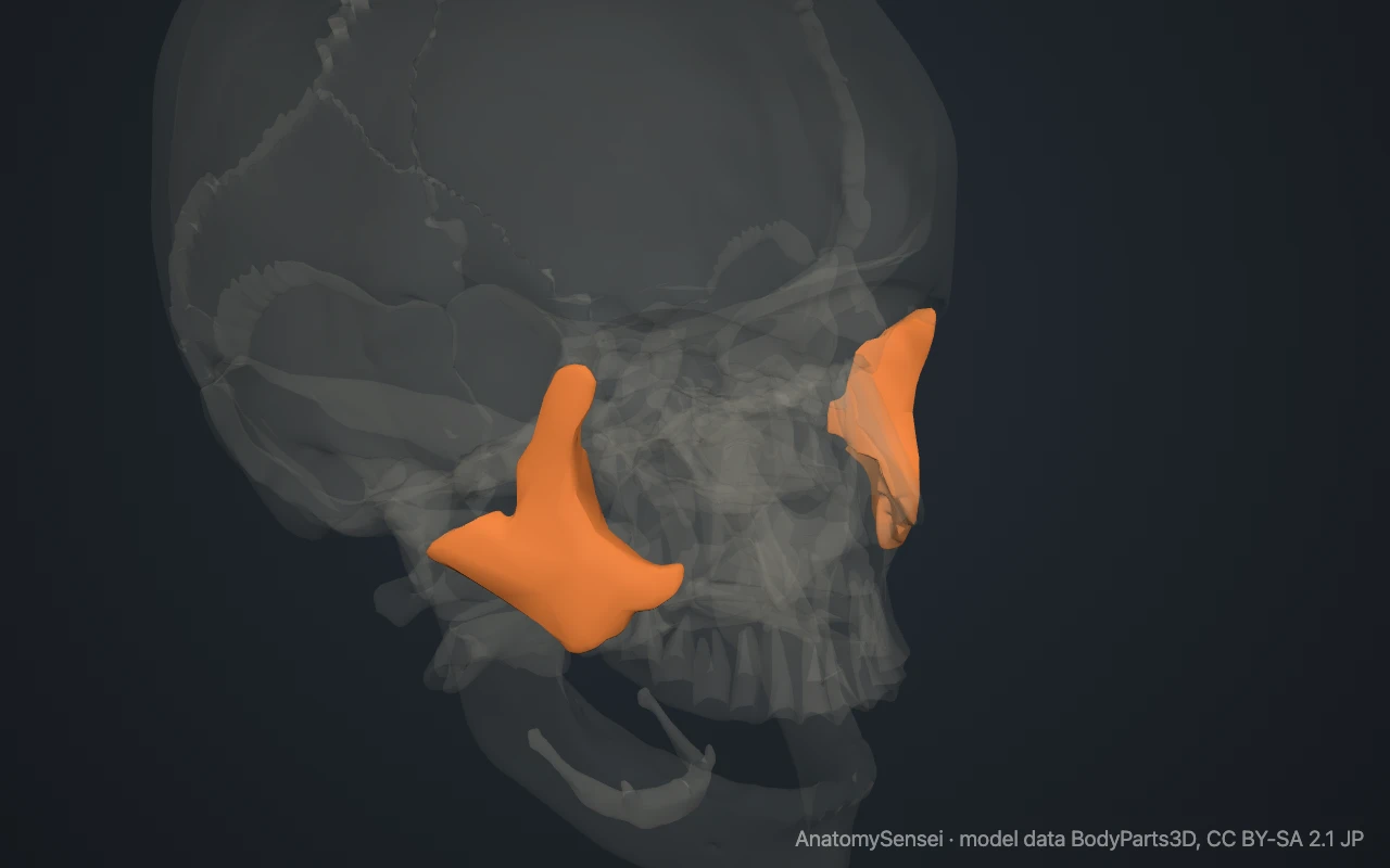
It is easy to feel on your own face. The firm, rounded prominence of the cheek below the outer corner of the eye is the body of the zygomatic bone. Slide a finger back from it toward the ear and you follow a narrow bar of bone. That bar is the zygomatic arch.
What are the parts of the zygomatic bone?
The bone has a body with three surfaces. The lateral surface faces the skin of the cheek. The orbital surface is smooth and faces into the eye socket. The temporal surface faces backward toward the side of the skull.
Two processes extend from the body. The frontal process runs upward along the outer edge of the orbit to meet the frontal bone. The temporal process runs backward to meet the temporal bone. The broad lower front part of the body, which rests on the maxilla, is sometimes called the maxillary process.
Small holes in the bone carry sensory nerves to the skin. The zygomaticofacial foramen opens on the cheek surface, and the zygomaticotemporal foramen opens on the temporal surface.
What does the zygomatic bone articulate with?
Each zygomatic bone joins four bones. It meets the maxilla below and in front, the frontal bone above, the temporal bone behind and the greater wing of the sphenoid bone inside the orbit.
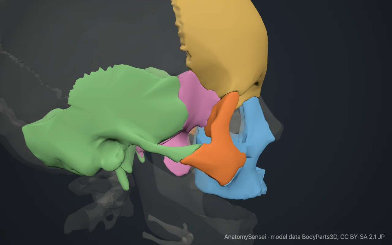
All four joints are sutures and do not move. The zygomatic bone does not touch the mandible, although the coronoid process of the mandible passes just inside the zygomatic arch.
What does the zygomatic bone form part of?
In the orbit, it forms the outer part of the rim, most of the outer wall and part of the floor. This outer rim is thick and protects the eye from blows that come from the side.
The temporal process joins a matching process of the temporal bone to make the zygomatic arch. The arch stands away from the side of the skull like a handle. The gap inside it lets the temporalis muscle pass down to the mandible.
What attaches to the zygomatic bone?
The masseter, the strongest chewing muscle, arises from the lower edge of the zygomatic bone and the zygomatic arch and runs down to the angle of the mandible.
Two muscles of facial expression, zygomaticus major and zygomaticus minor, arise from the cheek surface. They run to the corner of the mouth and the upper lip and pull them upward in a smile.
Common problems with the zygomatic bone
Because it projects, the cheekbone is often injured by a blow to the side of the face. A break may be limited to the zygomatic arch, which can be pushed inward.
A larger injury can break the bone free at its joints with the maxilla, frontal and temporal bones. This is called a zygomaticomaxillary complex fracture, or tripod fracture, and it can flatten the cheek and involve the floor of the orbit. This page describes anatomy only.
Common questions
Is the zygomatic bone the same as the cheekbone?
Yes. Zygomatic bone is the anatomical name for the cheekbone.
How many zygomatic bones are there?
Two, one on each side of the face.
Which bones make up the zygomatic arch?
The temporal process of the zygomatic bone and the zygomatic process of the temporal bone.
Is the zygomatic bone cranial or facial?
It is a facial bone, one of the 14 bones of the facial skeleton.
Is this 3D model of the zygomatic bone free?
Yes. The interactive 3D model on this page is free to view and use for study, with no account or download needed. Its geometry comes from the open BodyParts3D data set (CC BY-SA).
About this free 3D model of the zygomatic bone
The interactive 3D model on this page is free to use in your browser, with no sign-up. It is built from scan-based anatomy data, so every bone is a separate, correctly placed shape you can turn and inspect from any side.