
Vomer
Vomer
The vomer is a thin, flat, single bone in the midline of the nose. It forms the lower back part of the nasal septum, the wall that divides the nasal cavity into left and right halves.
Loading 3D model…
Free 3D model of the vomer. Whole skull. Drag to rotate, zoom with + and − (or Ctrl + scroll), click a bone to highlight and name it.
- Ethmoid bone
Key points
- The vomer is a single thin bone in the midline of the nose.
- It forms the lower back part of the nasal septum.
- It joins six bones: the sphenoid, the ethmoid, two maxillae and two palatine bones.
- Its free back edge separates the two rear openings of the nose.
What is the vomer?
The vomer is one of the two unpaired facial bones, the other being the mandible. It is a thin, flat plate of bone that stands upright in the midline of the nasal cavity.
Its name is Latin for a ploughshare, the blade of a plough, which describes its shape. Seen from the side it is a four-sided plate that is wider at the top back corner and tapers toward the front.
Where is the vomer?
It lies inside the nose, in the midline, running from the base of the skull above and behind down to the hard palate below. It occupies the lower and back part of the nasal septum.
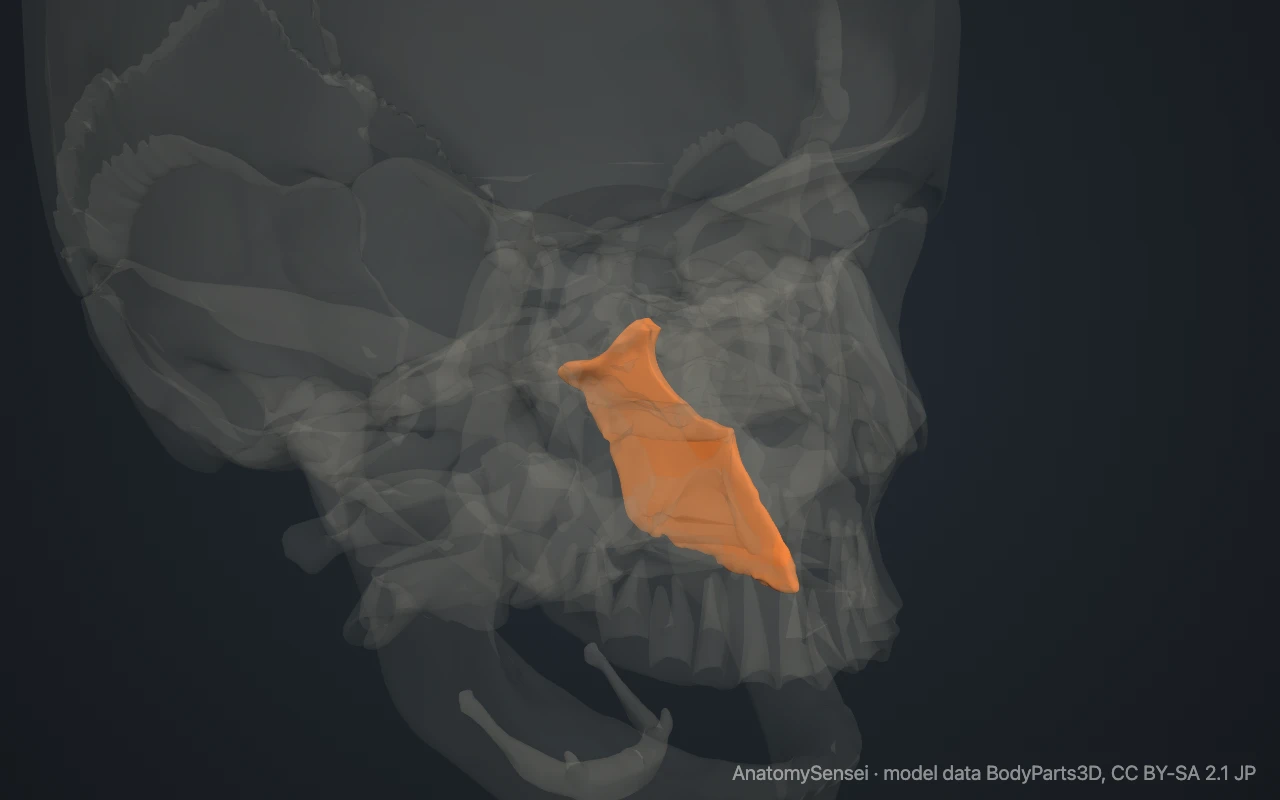
The vomer cannot be felt from outside. In a bare skull, its lower front edge can be seen by looking straight into the nasal opening, and its back edge can be seen from below, behind the hard palate. The model makes the other bones see-through to show it.
What are the parts of the vomer?
The vomer has two surfaces and four borders. The two surfaces, left and right, are flat and face the two halves of the nasal cavity. Each has a shallow groove that runs downward and forward.
The upper border is the thickest. It spreads into two small wings called the alae of the vomer, with a furrow between them. The lower border sits on the floor of the nose.
The front border slopes downward and forward. The back border is free and joins no other bone. It is smooth, slightly curved and thicker at the top.
What does the vomer articulate with?
The vomer joins six bones. Above, the furrow between its alae fits over a ridge on the underside of the body of the sphenoid bone. Below, it stands on the midline ridge formed by the two maxillae in front and the two palatine bones behind.
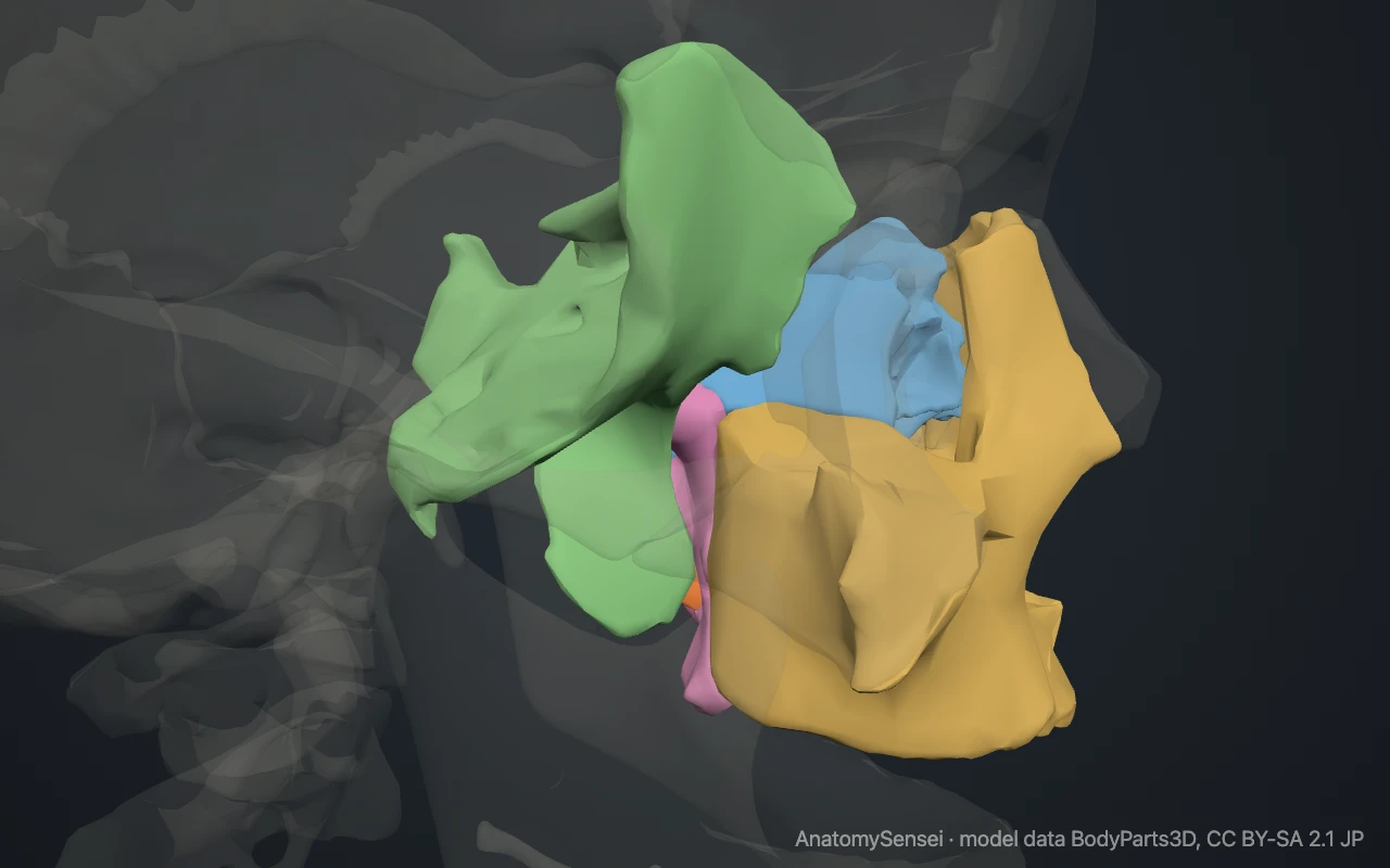
In front, its upper half meets the perpendicular plate of the ethmoid bone. Its lower half meets the septal cartilage of the nose, which is not bone and is not shown in the model.
What does the vomer form part of?
It forms the lower back part of the nasal septum. The septum has three main pieces. The perpendicular plate of the ethmoid forms the upper part, the vomer forms the lower back part, and the septal cartilage fills the gap in front.
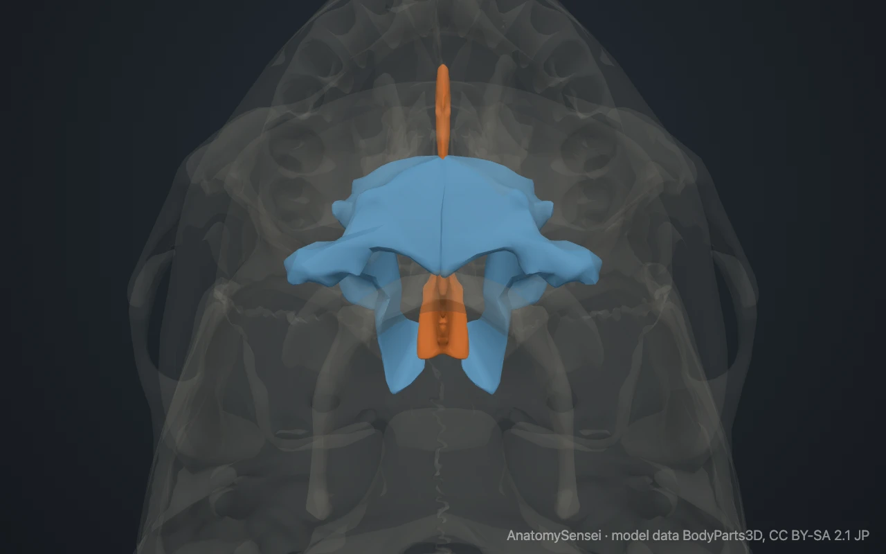
The free back border of the vomer separates the two choanae. These are the rear openings through which the nasal cavity leads into the throat.
What runs along the vomer?
No muscles attach to the vomer. Both of its surfaces are covered by the lining of the nose, which is firmly bound to the bone.
The groove on each surface carries the nasopalatine nerve and its accompanying vessels. They run downward and forward along the septum toward the incisive canal in the front of the hard palate.
What does the vomer do?
It divides the nasal cavity into two separate passages and helps to hold the septum upright. A straight septum gives each side of the nose a similar share of the airflow.
Common problems with the vomer
The vomer is often not perfectly in the midline. A septum that leans or bulges to one side is called a deviated septum. The bend may be in the cartilage, in the vomer, or at the joint between them, where a ridge of bone called a septal spur can form.
The vomer can also be damaged in fractures of the nose and middle of the face. This page describes anatomy only.
Common questions
Is the vomer a paired bone?
No. It is a single bone that lies in the midline.
Is the vomer a facial or a cranial bone?
It is a facial bone, one of the 14 bones of the facial skeleton.
Is the whole nasal septum made by the vomer?
No. The vomer forms the lower back part. The ethmoid bone forms the upper part and cartilage forms the front.
Why is it called the vomer?
Vomer is Latin for ploughshare. The bone is shaped like the blade of a plough.
Is this 3D model of the vomer free?
Yes. The interactive 3D model on this page is free to view and use for study, with no account or download needed. Its geometry comes from the open BodyParts3D data set (CC BY-SA).
About this free 3D model of the vomer
The interactive 3D model on this page is free to use in your browser, with no sign-up. It is built from scan-based anatomy data, so every bone is a separate, correctly placed shape you can turn and inspect from any side.