
Metacarpal bones
Ossa metacarpi
The metacarpals are the five long bones of the palm. They run from the carpal bones of the wrist to the knuckles, and each one carries a thumb or finger.
Loading 3D model…
Free 3D model of the metacarpal bones. Right hand. Drag to rotate, zoom with + and − (or Ctrl + scroll), click a bone to highlight and name it.
Key points
- There are five metacarpals, numbered from the thumb to the little finger.
- Each has a base at the wrist, a shaft, and a head that forms a knuckle.
- The first metacarpal is the shortest and most mobile. The second is usually the longest.
- The second and third are almost fixed, while the fourth and fifth move a little to cup the palm.
What are the metacarpal bones?
The metacarpals are the five bones that form the skeleton of the palm. The name means "beyond the carpus", because they lie just past the wrist bones. Together they are called the metacarpus.
They are small long bones. Like the femur or the humerus, each has a shaft with an enlarged end at either side, only on a much smaller scale.
Where are the metacarpal bones?
They lie between the carpal bones and the phalanges. Four of them fill the palm, and the first stands apart at the base of the thumb.
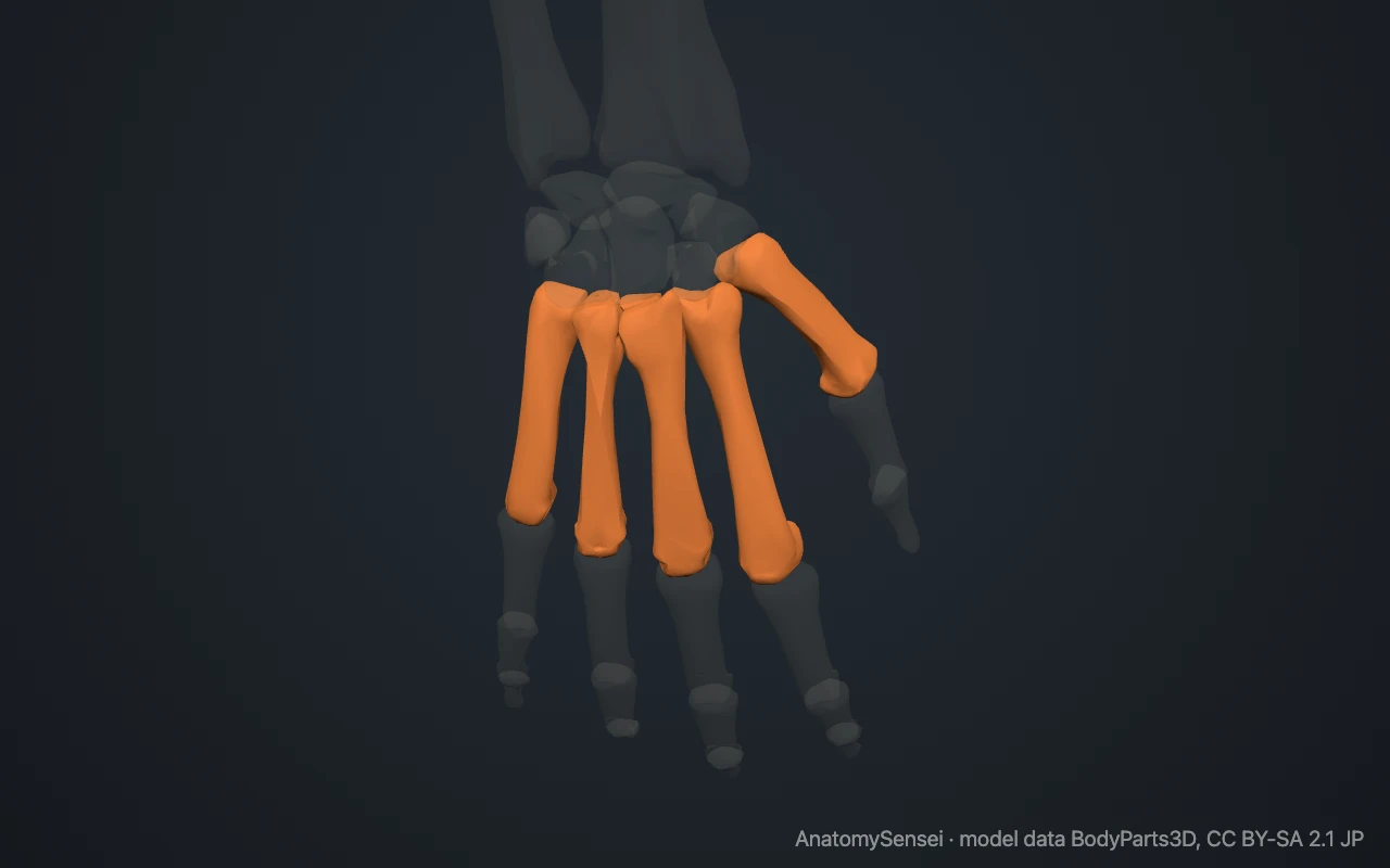
They are easy to feel on the back of your hand, where only skin and tendons cover them. Make a fist. The row of knuckles is the row of metacarpal heads. From each knuckle, a firm ridge can be traced back towards the wrist. That ridge is the shaft.
What are the parts of a metacarpal?
Each metacarpal has three parts. The base is the squared end at the wrist, with flat facets for the carpal bones and for the metacarpals beside it. The shaft is slightly curved, hollow towards the palm.
The head is the rounded far end. It fits into a shallow cup on the base of the proximal phalanx to form a knuckle joint. Just behind the head is a narrower neck, a frequent site of fracture.
How do the five metacarpals differ?
The first metacarpal belongs to the thumb. It is the shortest and thickest, and it is turned about a quarter turn so that the thumb faces across the palm. Its saddle-shaped base meets the trapezium and touches no other metacarpal.
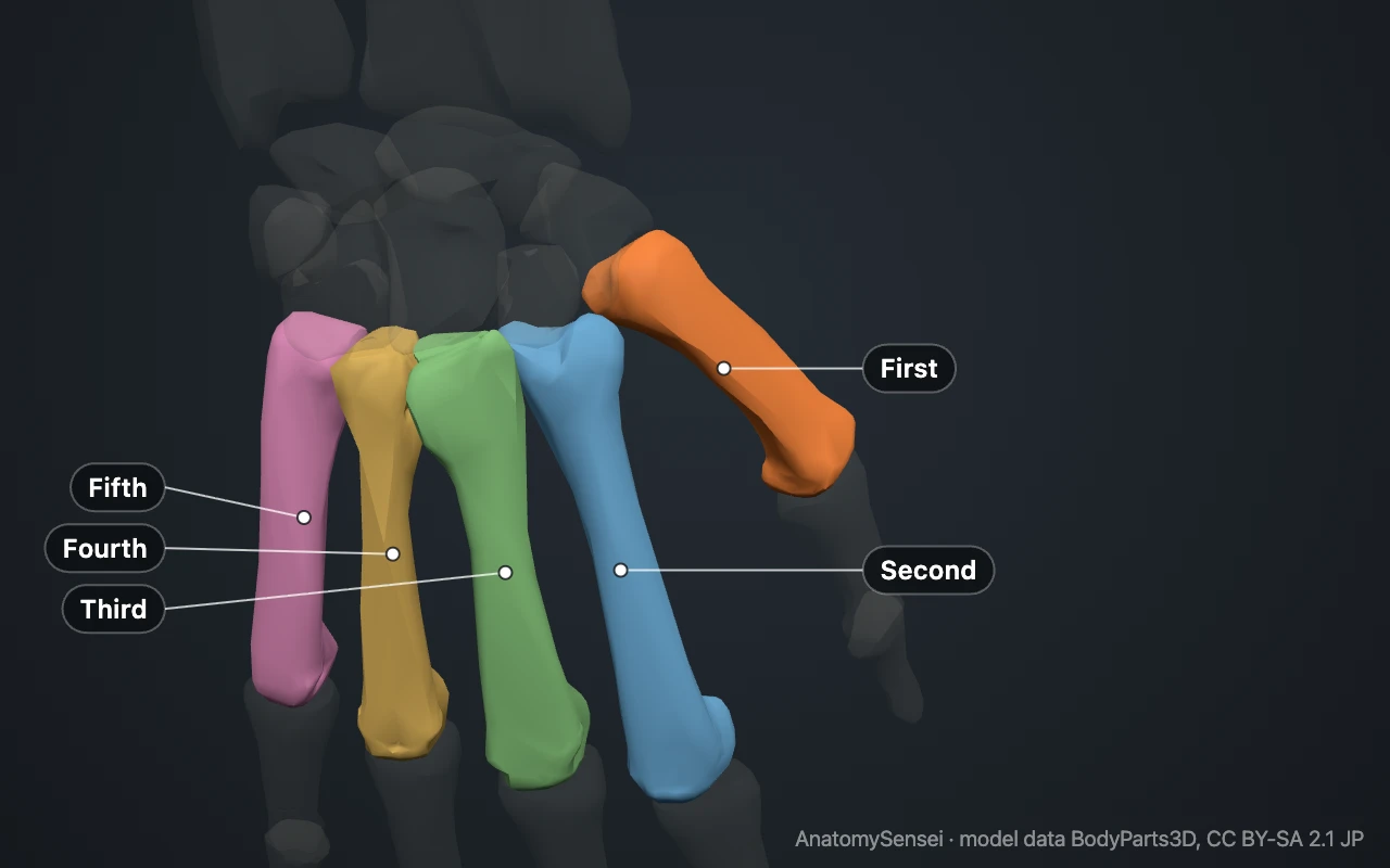
The second metacarpal, for the index finger, is usually the longest. Its forked base grips the trapezoid. The third metacarpal, for the middle finger, has a small styloid process, a point of bone that projects from the back of its base on the thumb side.
The fourth metacarpal, for the ring finger, is shorter and more slender. The fifth metacarpal, for the little finger, is the shortest of the four finger metacarpals and has a small bump on the outer side of its base for a tendon.
What do the metacarpals articulate with?
At the wrist, the bases meet the distal row of carpal bones. The first sits on the trapezium. The second meets mainly the trapezoid, the third the capitate, and the fourth and fifth the hamate. The bases of the second to fifth also touch each other.
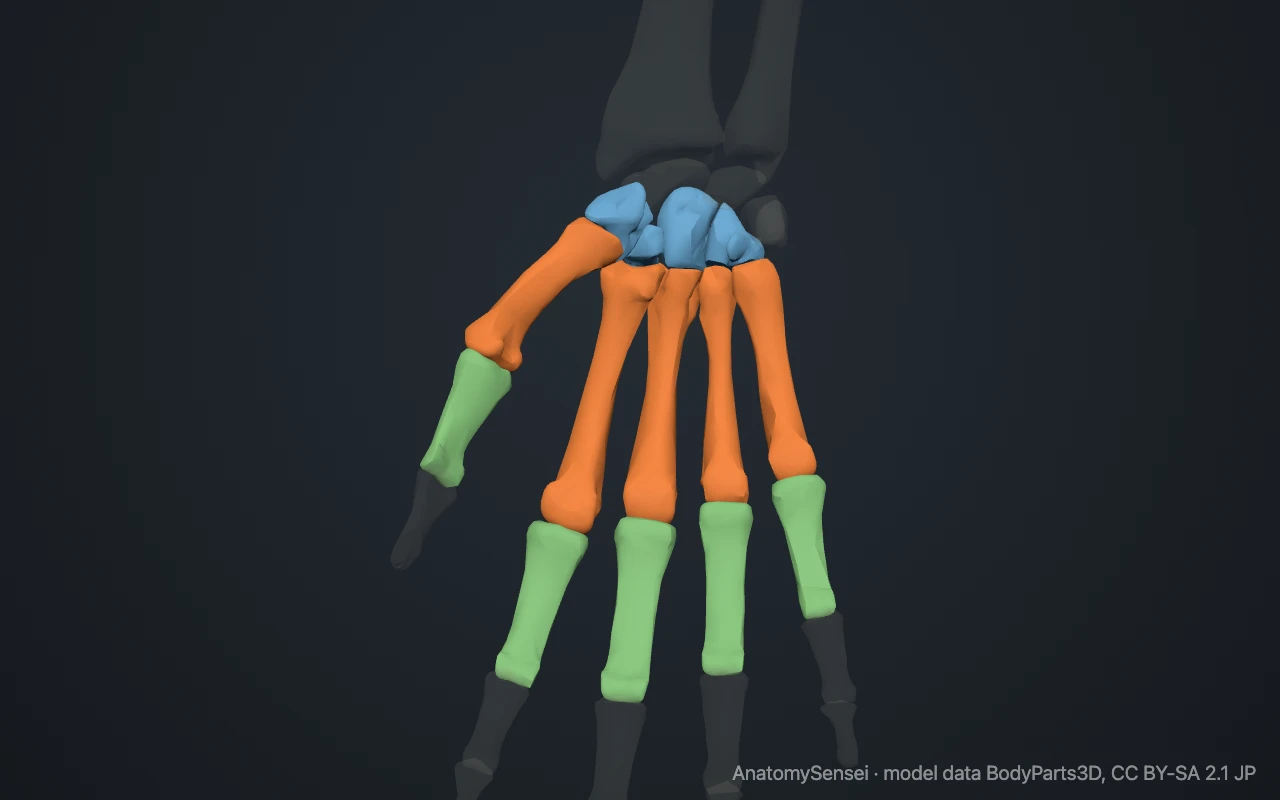
At the knuckles, each head meets the base of a proximal phalanx at a metacarpophalangeal joint. These joints let the fingers bend, straighten and spread.
What attaches to the metacarpals?
The sides of the shafts give origin to the interosseous muscles, the small muscles between the bones that spread the fingers and bring them together.
The main tendons that move the wrist end on the metacarpal bases. Flexor carpi radialis reaches the second, the two radial wrist extensors reach the second and third, and extensor carpi ulnaris reaches the fifth. The first metacarpal receives abductor pollicis longus at its base and opponens pollicis along its shaft.
What do the metacarpals do?
They form the frame of the palm and link the wrist to the fingers. The second and third metacarpals are held almost rigid on the carpus and make a fixed central pillar.
The others move around that pillar. The first swings freely to oppose the thumb. The fourth and fifth can tip forward slightly, which deepens the hollow of the palm and tightens a grip.
Common problems with the metacarpals
Metacarpal fractures are among the most common hand injuries. A break through the neck of the fifth metacarpal is known as a boxer's fracture, because it typically follows a punch against something hard.
A fracture through the base of the first metacarpal that enters the joint with the trapezium is called a Bennett fracture. Shaft fractures can follow a direct blow or a twisting injury of a finger. This page describes anatomy only.
Common questions
How many metacarpal bones are there?
Five in each hand, one for the thumb and one for each finger.
Are the knuckles part of the metacarpals?
Yes. The knuckles of a fist are the heads of the metacarpals.
Which metacarpal is the longest?
Usually the second, the metacarpal of the index finger.
Which metacarpal has a styloid process?
The third. A small point of bone projects from the back of its base.
What is the difference between metacarpals and metatarsals?
Metacarpals are the five bones of the palm. Metatarsals are the five matching bones of the foot.
Is this 3D model of the metacarpal bones free?
Yes. The interactive 3D model on this page is free to view and use for study, with no account or download needed. Its geometry comes from the open BodyParts3D data set (CC BY-SA).
About this free 3D model of the metacarpal bones
The interactive 3D model on this page is free to use in your browser, with no sign-up. It is built from scan-based anatomy data, so every bone is a separate, correctly placed shape you can turn and inspect from any side.