
Lunate bone
Os lunatum
The lunate is a crescent-shaped carpal bone in the middle of the proximal row of the wrist. It sits between the radius and the capitate and is the carpal bone most often dislocated.
Loading 3D model…
Free 3D model of the lunate bone. Right hand. Drag to rotate, zoom with + and − (or Ctrl + scroll), click a bone to highlight and name it.
Key points
- The lunate is the central bone of the proximal row of the wrist.
- It is shaped like a crescent moon, with a hollow that holds the head of the capitate.
- With the scaphoid, it forms the carpal side of the wrist joint with the radius.
- It is the most commonly dislocated carpal bone.
What is the lunate bone?
The lunate is one of the eight carpal bones. Its name comes from the Latin luna, the moon, because from the side it looks like a crescent. It is sometimes called the semilunar bone.
It is the middle bone of the proximal row, with the scaphoid on its thumb side and the triquetrum on its little finger side.
Where is the lunate bone?
It lies in the centre of the wrist, just beyond the end of the radius and in line with the middle finger. The capitate sits directly beyond it.
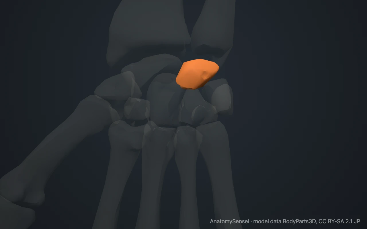
To find it, place a fingertip on the back of your wrist in line with the middle finger, just past the edge of the radius. Bend the wrist forward. The rounded bone that pushes up under the finger is the lunate.
What does the lunate look like?
The lunate has a convex surface facing the forearm and a deeply concave surface facing the hand. The convex side fits against the radius. The concave side is the hollow of the crescent.
The bone is wider on the palm side than on the back. Its palm and back surfaces are small and rough, for ligaments. The sides are flat and smooth where they meet the scaphoid and triquetrum.
What does the lunate articulate with?
On the forearm side the lunate meets the radius, and part of it faces the cartilage disc at the end of the ulna. With the scaphoid it forms the carpal half of the wrist joint.
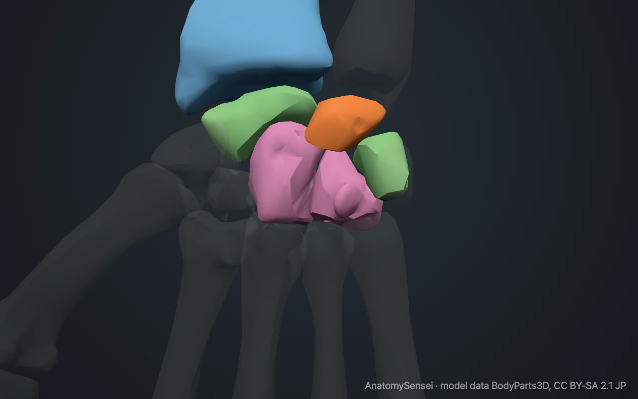
On the hand side its hollow receives the head of the capitate. It usually also has a narrow contact with the hamate. On either side it meets the scaphoid and the triquetrum, to which it is bound by short, strong ligaments.
What attaches to the lunate?
No muscles or tendons attach to the lunate. It is held in place only by ligaments and by the shape of the bones around it.
The most important of these are the scapholunate and lunotriquetral ligaments, which tie it to its neighbours in the row. Just in front of the lunate lies the carpal tunnel, with the median nerve and the tendons that bend the fingers.
What does the lunate do?
The lunate is the central link between the forearm and the hand. Force from the hand passes from the capitate into the lunate and on to the radius.
It also moves with the wrist. Because nothing pulls on it directly, it is pushed and guided by its neighbours, tipping forward and back as the wrist bends. For this reason it is described as an intercalated segment, a middle piece controlled from both ends.
Common problems with the lunate
The lunate is the carpal bone most often dislocated. After a hard fall on a bent-back wrist it can be pushed forward into the carpal tunnel, where it may press on the median nerve.
In Kienböck disease, the blood supply to the lunate fails and the bone gradually collapses. A tear of the ligament between the scaphoid and the lunate lets the two bones drift apart and is a common cause of wrist instability. This page describes anatomy only.
Common questions
Why is it called the lunate?
From the Latin for moon. Seen from the side, the bone is shaped like a crescent.
Do any muscles attach to the lunate?
No. It is held only by ligaments and by the surrounding bones.
Which bones touch the lunate?
The radius, scaphoid, triquetrum and capitate, and usually the hamate.
Is the lunate in the proximal or distal row?
The proximal row, between the scaphoid and the triquetrum.
Is this 3D model of the lunate bone free?
Yes. The interactive 3D model on this page is free to view and use for study, with no account or download needed. Its geometry comes from the open BodyParts3D data set (CC BY-SA).
About this free 3D model of the lunate bone
The interactive 3D model on this page is free to use in your browser, with no sign-up. It is built from scan-based anatomy data, so every bone is a separate, correctly placed shape you can turn and inspect from any side.