
Medial cuneiform
Os cuneiforme mediale
The medial cuneiform is the largest of the three cuneiform bones. It sits on the inner side of the midfoot between the navicular and the first metatarsal.
Loading 3D model…
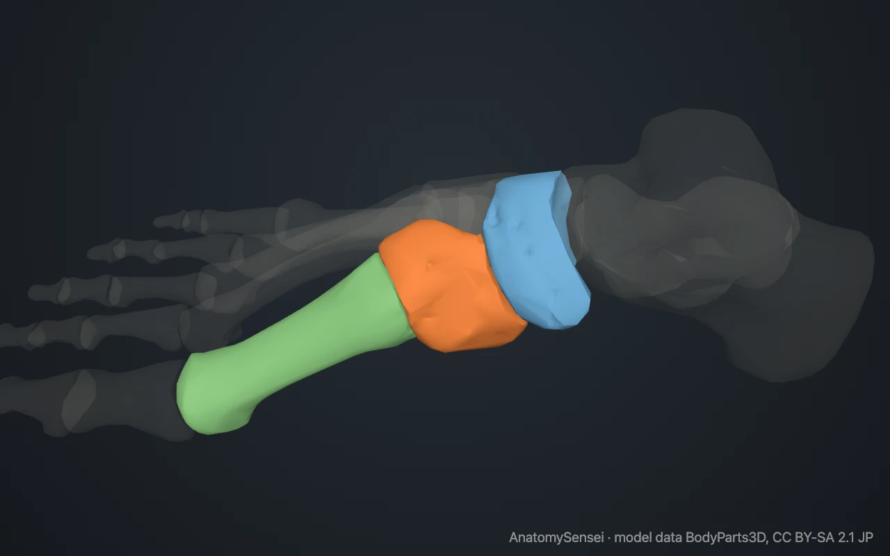
Free 3D model of the medial cuneiform. Right foot. Drag to rotate, zoom with + and − (or Ctrl + scroll), click a bone to highlight and name it.
Key points
- The medial cuneiform is the largest of the three cuneiform bones.
- It sits on the inner side of the midfoot, behind the first metatarsal.
- Two tendons that support the arch attach to it.
- It is also called the first cuneiform.
What is the medial cuneiform?
The medial cuneiform is the innermost and largest of the three cuneiform bones. It is one of the seven tarsals and part of the midfoot.
Where is the medial cuneiform?
It lies on the inner side of the foot, between the navicular and the first metatarsal. It can be felt just behind the base of the first metatarsal.
What does the medial cuneiform articulate with?
It meets four bones: the navicular behind, the first metatarsal in front, and on its outer side the intermediate cuneiform and the base of the second metatarsal.
Shape of the medial cuneiform
It is a wedge with its broad edge underneath, the reverse of the other two cuneiforms. Its front surface is kidney-shaped to match the base of the first metatarsal.
What attaches to the medial cuneiform?
Tibialis anterior attaches to its inner side and fibularis longus to its underside. Both also attach to the neighbouring base of the first metatarsal, and together they steady the inner arch.
A strong ligament runs from this bone to the base of the second metatarsal and is the main tie between the inner and middle parts of the midfoot.
What does the medial cuneiform do?
It is the base of the inner column of the foot. Load arriving from the navicular passes through it to the first metatarsal and on to the big toe.
The joint in front of it, with the first metatarsal, allows a little movement. How much it moves affects how stable the first metatarsal is, which is why this joint gets attention in bunion surgery.
Common problems with the medial cuneiform
In some people the bone forms in two pieces, an upper and a lower. This is a normal variant. The bone is also involved in Lisfranc injuries of the midfoot. This page describes anatomy only.
Common questions
Where is the medial cuneiform?
On the inner side of the midfoot, behind the first metatarsal.
What attaches to the medial cuneiform?
The tibialis anterior and fibularis longus tendons.
Which is the largest cuneiform?
The medial cuneiform.
Is this 3D model of the medial cuneiform free?
Yes. The interactive 3D model on this page is free to view and use for study, with no account or download needed. Its geometry comes from the open BodyParts3D data set (CC BY-SA).
About this free 3D model of the medial cuneiform
The interactive 3D model on this page is free to use in your browser, with no sign-up. It is built from scan-based anatomy data, so every bone is a separate, correctly placed shape you can turn and inspect from any side.