
Tarsal bones
Ossa tarsi
The tarsal bones are the seven bones of the ankle and midfoot: the talus, calcaneus, navicular, cuboid and the medial, intermediate and lateral cuneiforms.
Loading 3D model…
Free 3D model of the tarsal bones. Right foot. Drag to rotate, zoom with + and − (or Ctrl + scroll), click a bone to highlight and name it.
Key points
- There are seven tarsal bones in each foot.
- Two form the hindfoot (talus, calcaneus) and five form the midfoot.
- The talus joins the foot to the leg; the calcaneus is the heel.
- They are the foot's counterpart to the eight carpal bones of the wrist.
What are the tarsal bones?
The tarsal bones are the seven bones of the ankle and the back half of the foot. They sit between the lower ends of the leg bones and the bases of the metatarsals. Together they are called the tarsus.
They are short, thick bones built to carry load. Every step passes body weight through them.
The seven tarsal bones
The talus is the top bone and forms the ankle joint with the tibia and fibula. The calcaneus lies under it and is the heel. The navicular sits in front of the talus on the inner side.
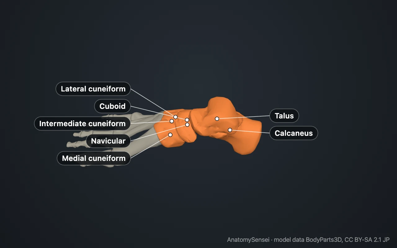
The medial, intermediate and lateral cuneiforms line up in front of the navicular, one behind each of the first three metatarsals. The cuboid is on the outer side, between the calcaneus and the fourth and fifth metatarsals.
Hindfoot and midfoot
The talus and calcaneus make up the hindfoot. They are the largest two and take load first. The other five make up the midfoot, a compact block that forms the top of the arch.
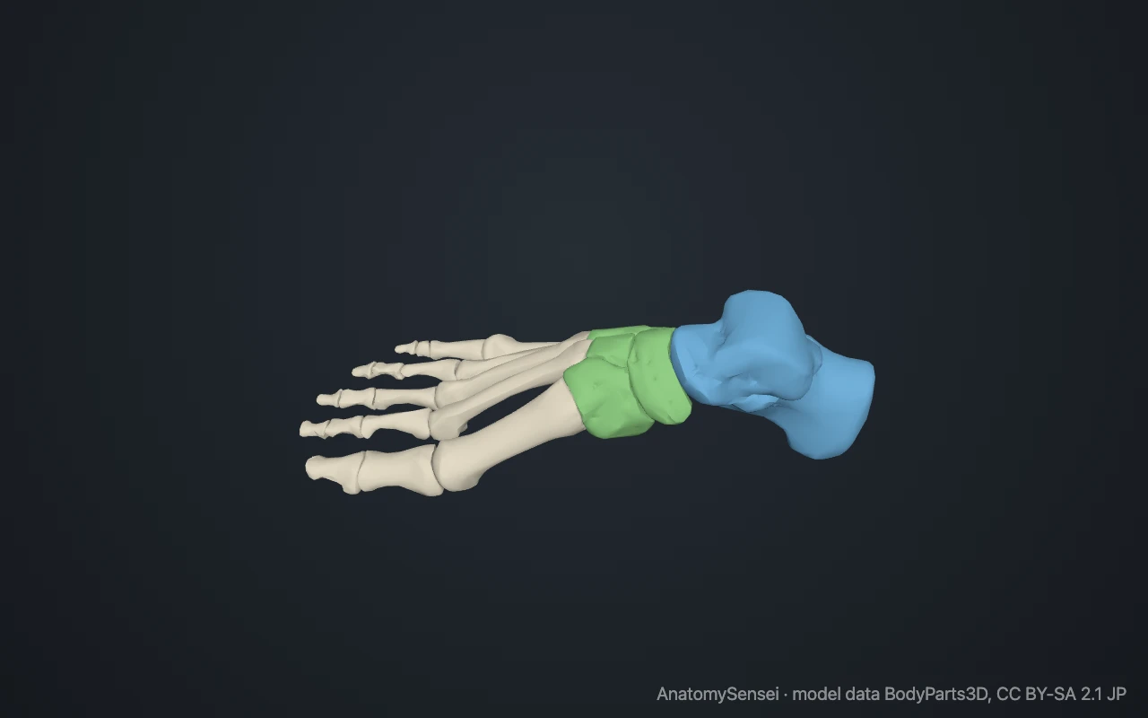
Three sets of joints link them. The subtalar joint lies between talus and calcaneus. The transverse tarsal joint separates hindfoot from midfoot. The tarsometatarsal joints connect the midfoot to the metatarsals.
What do the tarsal bones do?
They pass weight from the leg to the ground. The talus receives it and shares it between the heel behind and the forefoot in front.
They also give the foot its arched shape. The wedge form of the cuneiforms and the way the navicular locks onto the talus hold the arch up even before muscles act.
How to remember the tarsal bones
A common memory aid is "Tiger Cubs Need MILC": Talus, Calcaneus, Navicular, then the Medial, Intermediate and Lateral cuneiforms, and the Cuboid.
Common problems with the tarsal bones
The calcaneus is the tarsal bone most often broken, usually in a fall from a height onto the heels. Fractures of the talus are less common but matter because of its fragile blood supply.
A tarsal coalition is an abnormal bridge of bone or cartilage joining two tarsal bones, most often the calcaneus and navicular or the talus and calcaneus. It can cause a stiff, flat foot in adolescence. This page describes anatomy only; pain or stiffness needs a clinician.
Common questions
How many tarsal bones are there?
Seven in each foot.
Which tarsal bone is the largest?
The calcaneus. The talus is second.
Which tarsal bone articulates with the tibia?
The talus. Its domed upper surface fits between the tibia and fibula.
What is the difference between tarsals and carpals?
Tarsals are in the foot and there are seven. Carpals are in the wrist and there are eight.
Is this 3D model of the tarsal bones free?
Yes. The interactive 3D model on this page is free to view and use for study, with no account or download needed. Its geometry comes from the open BodyParts3D data set (CC BY-SA).
About this free 3D model of the tarsal bones
The interactive 3D model on this page is free to use in your browser, with no sign-up. It is built from scan-based anatomy data, so every bone is a separate, correctly placed shape you can turn and inspect from any side.