
Talus
Os tali
The talus is the tarsal bone that sits between the shin bones and the heel bone. It forms the lower half of the ankle joint and passes body weight from the leg into the foot.
Loading 3D model…
Free 3D model of the talus. Right foot. Drag to rotate, zoom with + and − (or Ctrl + scroll), click a bone to highlight and name it.
Key points
- The talus is the bone that joins the foot to the leg.
- It sits between the tibia and fibula above and the heel bone below.
- No muscles attach to it, and most of its surface is joint cartilage.
- Its blood supply is limited, which makes some fractures slow to heal.
What is the talus?
The talus is the tarsal bone that links the leg to the foot. All of the body's weight passes through it with each step. It is the second largest tarsal bone, after the calcaneus.
Where is the talus?
It sits at the top of the foot, directly under the shin bone and on top of the heel bone. It lies deep between the two ankle bumps, so most of it cannot be felt.
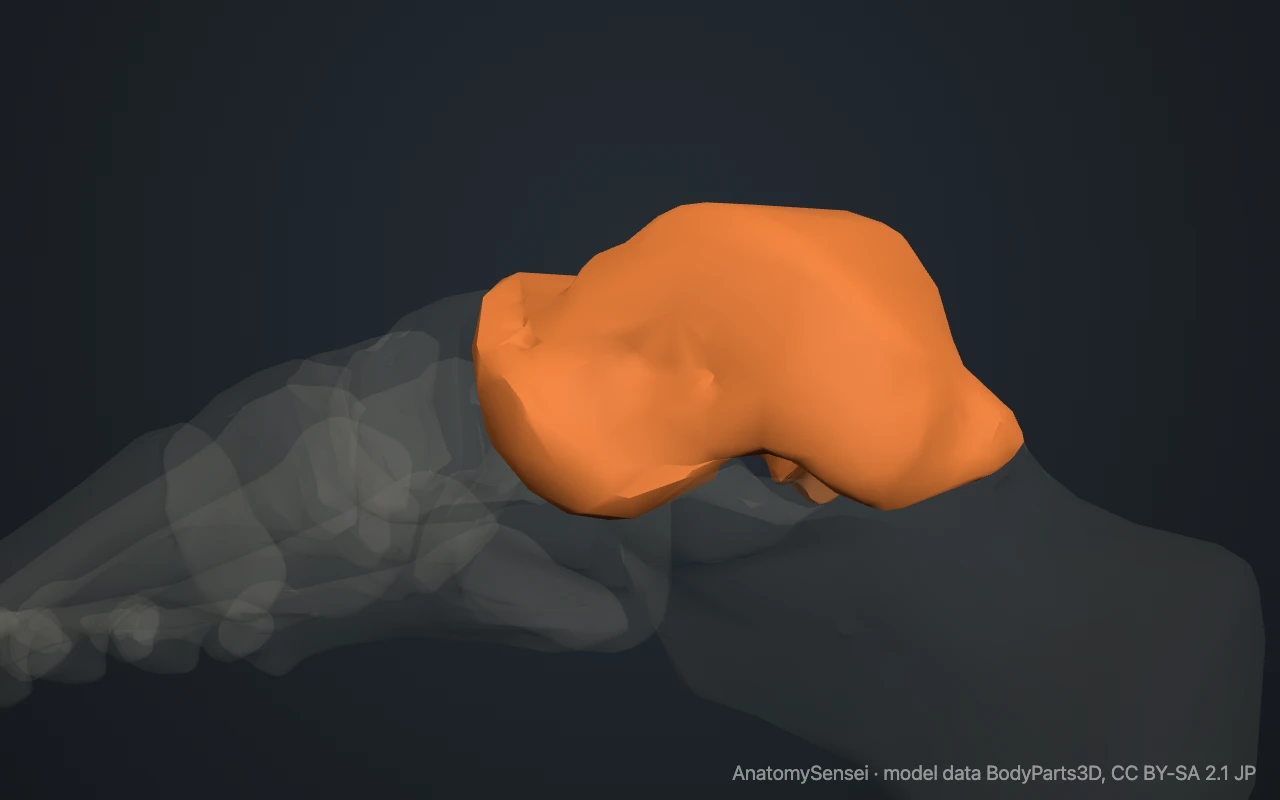
A small part of its front can be felt just in front of the ankle when the foot is pointed down and turned in.
Parts of the talus
The talus has three parts. The body is the large rear portion. Its upper surface is a smooth dome called the trochlea, which fits into the ankle socket. The neck is the narrower section in front. The head is the rounded front end.
At the back, a small posterior process has two bumps with a groove between them for the tendon that bends the big toe.
What does the talus articulate with?
It meets four bones. Above and to the sides, the trochlea fits between the tibia and fibula to form the ankle joint. Below, it rests on the calcaneus at the subtalar joint. In front, its head fits into the navicular.
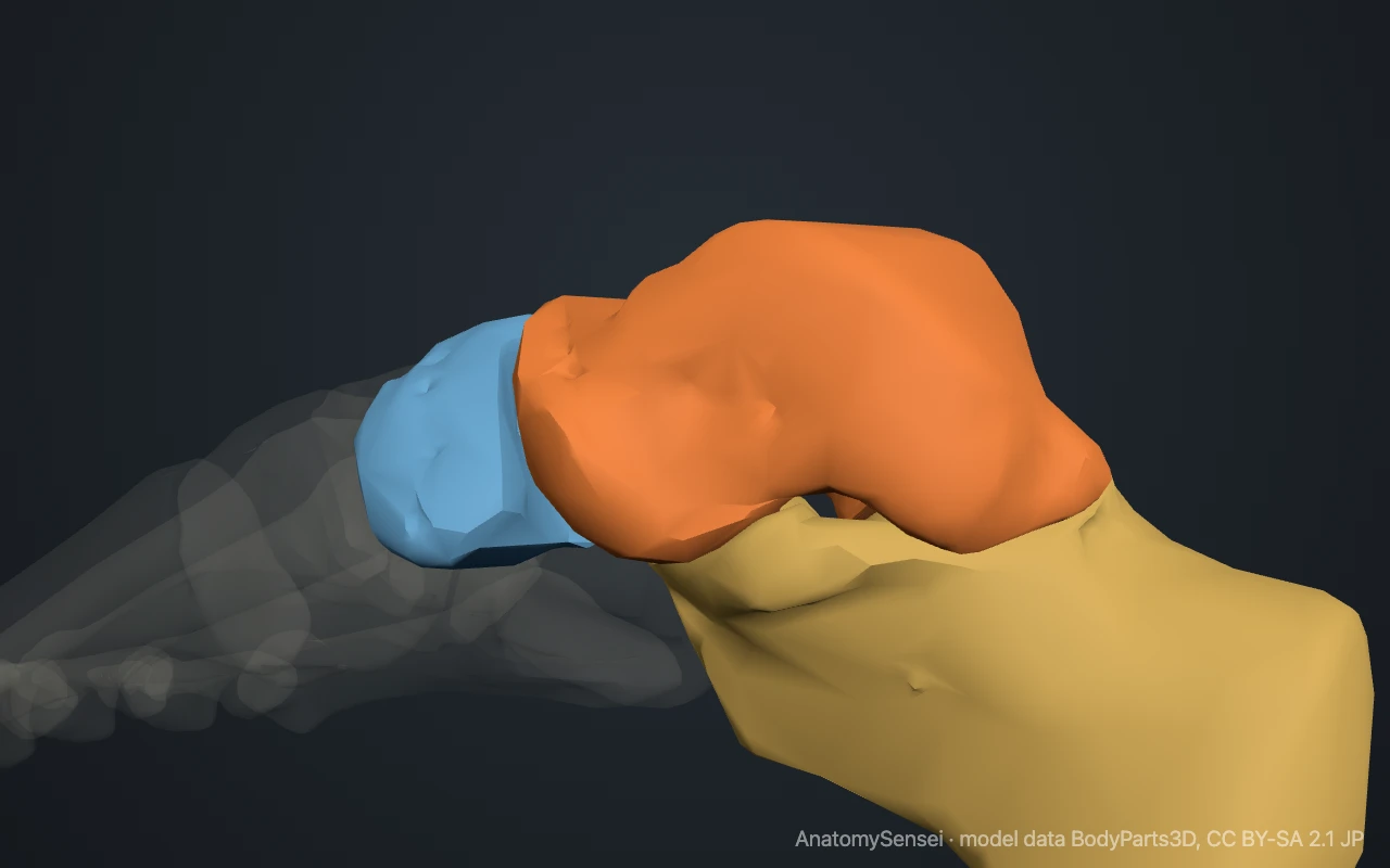
The ankle joint moves the foot up and down. The subtalar joint tilts it in and out.
Why the talus is unusual
No muscles attach to the talus. It is moved entirely by the bones around it. About two thirds of its surface is covered in joint cartilage.
That leaves few places for blood vessels to enter, and much of the supply runs backward from the neck into the body. A fracture through the neck can cut it off, so part of the bone may fail to heal.
Common problems with the talus
Talus fractures are uncommon and usually follow high-energy injuries such as falls and road accidents. Damage to the cartilage on the dome can follow a bad ankle sprain.
Some people have a small extra bone behind the talus called an os trigonum. It is a normal variant that occasionally gets pinched when the foot is pointed. This page describes anatomy only.
Common questions
Is the talus part of the ankle or the foot?
Both. It is a foot bone, and its upper surface forms the lower half of the ankle joint.
Can you feel your talus?
Mostly no. It sits deep between the ankle bumps. Part of its front can be felt just in front of the ankle.
What type of bone is the talus?
A short bone, one of the seven tarsals.
What is another name for the talus?
The ankle bone. An older name is astragalus.
Is this 3D model of the talus free?
Yes. The interactive 3D model on this page is free to view and use for study, with no account or download needed. Its geometry comes from the open BodyParts3D data set (CC BY-SA).
About this free 3D model of the talus
The interactive 3D model on this page is free to use in your browser, with no sign-up. It is built from scan-based anatomy data, so every bone is a separate, correctly placed shape you can turn and inspect from any side.