
Cuneiform bones
Ossa cuneiformia
The cuneiforms are three wedge-shaped bones in the midfoot. They sit in a row between the navicular and the first three metatarsals and are named medial, intermediate and lateral.
Loading 3D model…
Free 3D model of the cuneiform bones. Right foot. Drag to rotate, zoom with + and − (or Ctrl + scroll), click a bone to highlight and name it.
Key points
- There are three cuneiform bones in each foot: medial, intermediate and lateral.
- They sit in a row between the navicular and the first three metatarsals.
- Their wedge shape builds the arch across the foot.
- The medial is the largest and the intermediate the smallest.
What are the cuneiform bones?
The cuneiforms are three wedge-shaped tarsal bones in the midfoot. The name comes from the Latin cuneus, a wedge.
Medial, intermediate and lateral
They are named by position. The medial cuneiform is on the inner side and is the largest. The intermediate cuneiform is in the middle and is the smallest. The lateral cuneiform is on the outer side, next to the cuboid.
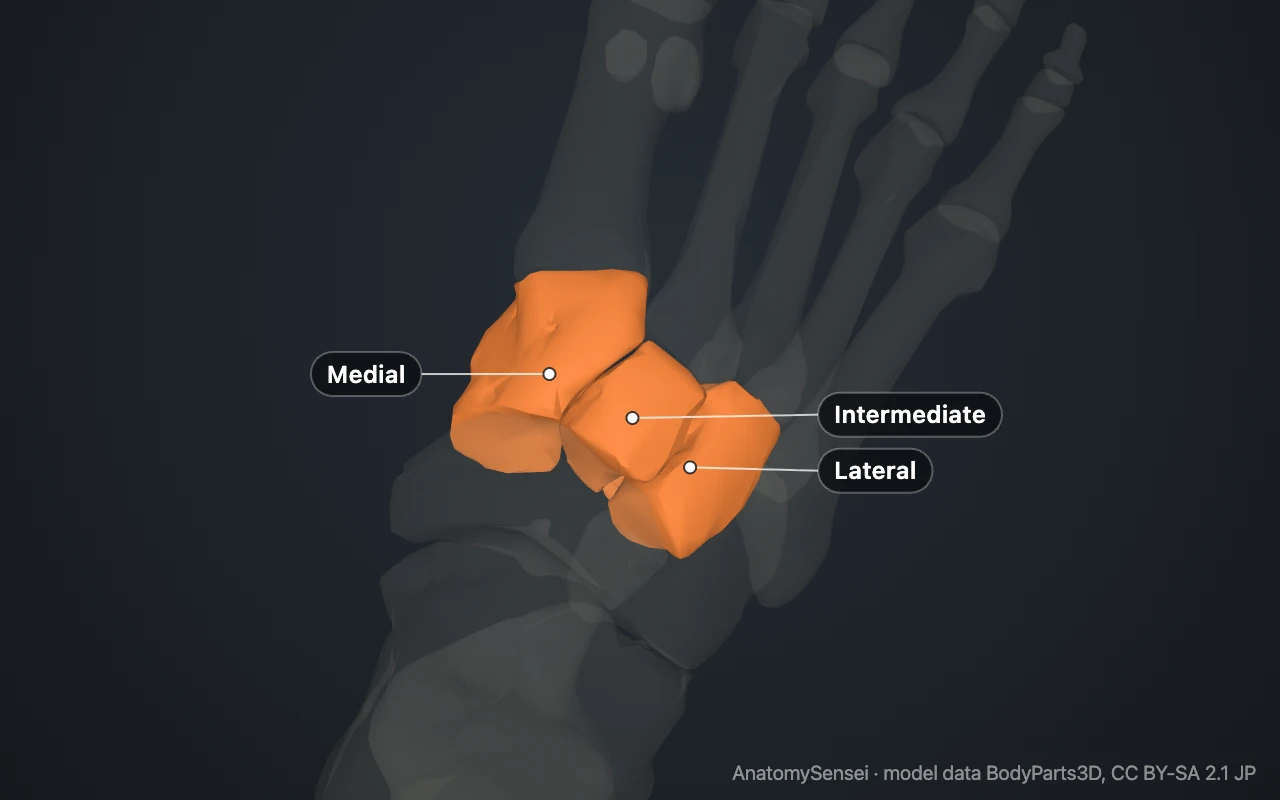
They are also called the first, second and third cuneiforms.
What do the cuneiforms articulate with?
Behind, all three meet the navicular. In front, each lines up with one metatarsal: medial with the first, intermediate with the second, lateral with the third.
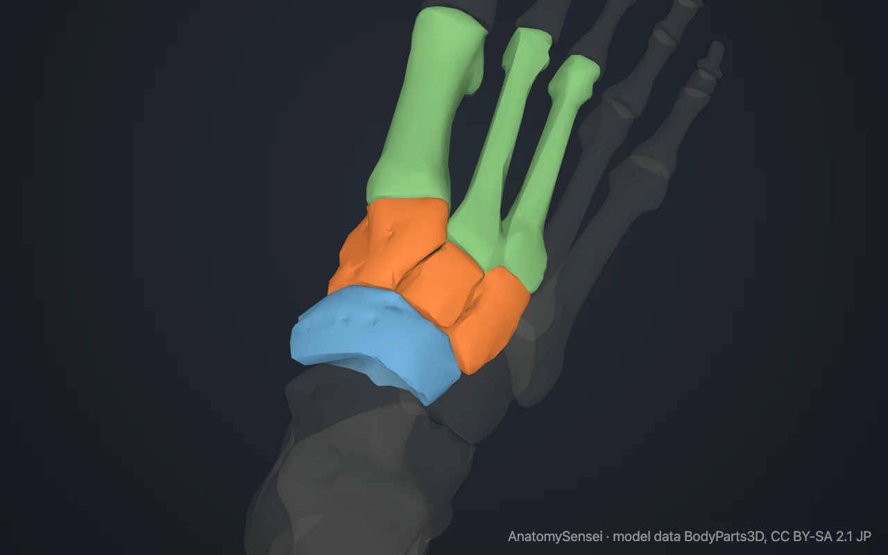
The lateral cuneiform also touches the cuboid on its outer side. Neighbouring cuneiforms touch each other.
Why the second metatarsal is locked in place
The intermediate cuneiform is shorter than the other two. That leaves a slot between the medial and lateral cuneiforms, and the base of the second metatarsal sits in it.
The slot grips the second metatarsal and makes it the most stable ray of the foot. This arrangement is the keystone of the midfoot.
What do the cuneiforms do?
Their wedge shape, narrow underneath for the intermediate and lateral, forms the arch that runs across the foot. They also pass load from the navicular forward to the metatarsals.
Two tendons that support the arch, tibialis anterior and fibularis longus, attach to the medial cuneiform.
Common problems with the cuneiforms
A break of a single cuneiform is rare. They are more often involved in a Lisfranc injury, in which the joints between the cuneiforms and the metatarsals are disrupted.
Arthritis of the midfoot can produce a bony ridge on top of the foot over these joints. This page describes anatomy only.
Common questions
How many cuneiform bones are there?
Three in each foot.
Which cuneiform is the largest?
The medial cuneiform. The intermediate is the smallest.
Why are they called cuneiform?
From the Latin for wedge, after their shape.
Where are the cuneiform bones?
In the midfoot, behind the first three metatarsals and in front of the navicular.
Is this 3D model of the cuneiform bones free?
Yes. The interactive 3D model on this page is free to view and use for study, with no account or download needed. Its geometry comes from the open BodyParts3D data set (CC BY-SA).
About this free 3D model of the cuneiform bones
The interactive 3D model on this page is free to use in your browser, with no sign-up. It is built from scan-based anatomy data, so every bone is a separate, correctly placed shape you can turn and inspect from any side.