
Intermediate cuneiform
Os cuneiforme intermedium
The intermediate cuneiform is the smallest of the three cuneiform bones. It sits between the medial and lateral cuneiforms and carries the base of the second metatarsal.
Loading 3D model…
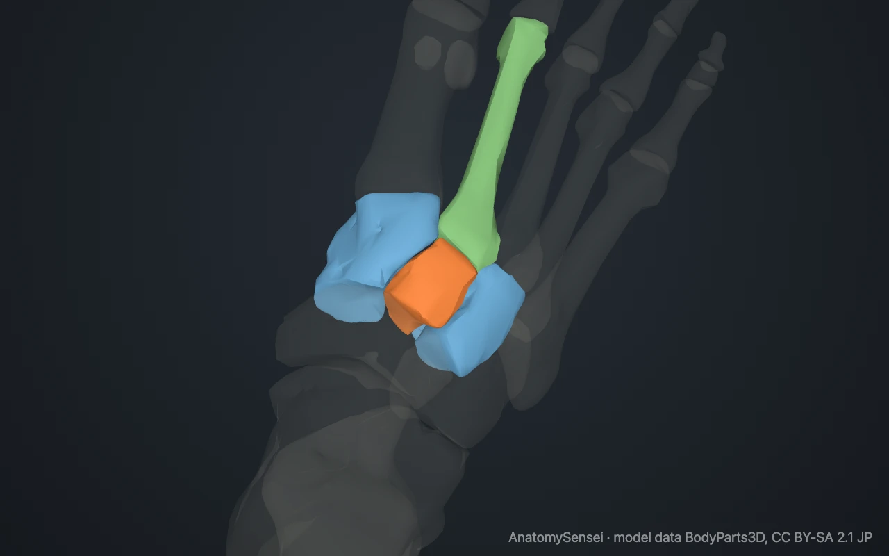
Free 3D model of the intermediate cuneiform. Right foot. Drag to rotate, zoom with + and − (or Ctrl + scroll), click a bone to highlight and name it.
Key points
- The intermediate cuneiform is the smallest of the three cuneiforms.
- It sits between the medial and lateral cuneiforms.
- It carries the base of the second metatarsal.
- Because it is short, the second metatarsal is locked in beside it.
What is the intermediate cuneiform?
The intermediate cuneiform is the middle and smallest of the three cuneiform bones. It is also called the second or middle cuneiform.
Where is the intermediate cuneiform?
It lies in the middle of the midfoot, directly behind the second metatarsal, with a cuneiform on either side and the navicular behind.
What does the intermediate cuneiform articulate with?
It meets four bones: the navicular behind, the second metatarsal in front, and the medial and lateral cuneiforms on either side.
Why its size matters
It is shorter from back to front than the cuneiforms beside it. The gap this leaves is filled by the base of the second metatarsal, which is gripped between the medial and lateral cuneiforms.
It is wedge-shaped with its narrow edge down, which helps form the arch across the foot.
Can you feel the intermediate cuneiform?
Only roughly. Trace the ridge of the second metatarsal back from the second toe along the top of your foot. Where the ridge ends, in the middle of the instep, you are over the intermediate cuneiform. It sits near the highest point of the arch that crosses the foot.
What attaches to the intermediate cuneiform?
Very little muscle. A slip of the tibialis posterior tendon reaches its underside. Its main connections are ligaments: short, strong bands tie it to the navicular behind, to the cuneiform on each side and to the second metatarsal in front.
Those ligaments, more than any muscle, are what keep the middle of the foot rigid.
Common problems with the intermediate cuneiform
It is rarely injured on its own. Arthritis of the joint in front of it, at the base of the second metatarsal, is a common cause of a bony ridge on top of the foot. This page describes anatomy only.
Common questions
Where is the intermediate cuneiform?
In the middle of the midfoot, behind the second metatarsal.
Which cuneiform is the smallest?
The intermediate cuneiform.
What is another name for the intermediate cuneiform?
The second or middle cuneiform.
Is this 3D model of the intermediate cuneiform free?
Yes. The interactive 3D model on this page is free to view and use for study, with no account or download needed. Its geometry comes from the open BodyParts3D data set (CC BY-SA).
About this free 3D model of the intermediate cuneiform
The interactive 3D model on this page is free to use in your browser, with no sign-up. It is built from scan-based anatomy data, so every bone is a separate, correctly placed shape you can turn and inspect from any side.