
Tibia (shin bone)
Tibia
The tibia is the shin bone. It is the larger of the two bones of the leg, lies on the inner side, and carries the weight of the body from the knee to the ankle.
Loading 3D model…
Free 3D model of the tibia. Right leg. Drag to rotate, zoom with + and − (or Ctrl + scroll), click a bone to highlight and name it.
Key points
- The tibia is the shin bone, on the inner side of the leg.
- It is the second longest bone in the body and the weight-bearing bone of the leg.
- Its flat top meets the femur at the knee and its lower end meets the talus at the ankle.
- Its lower end forms the medial malleolus, the bump on the inner side of the ankle.
What is the tibia?
The tibia, or shin bone, is the larger of the two leg bones. Its partner, the slender fibula, lies beside it on the outer side.
After the femur it is the second longest bone in the body. It takes nearly all of the load that passes from the knee to the foot.
Where is the tibia?
It runs from the knee to the ankle on the inner side of the leg. Unlike the femur, much of it lies directly under the skin.
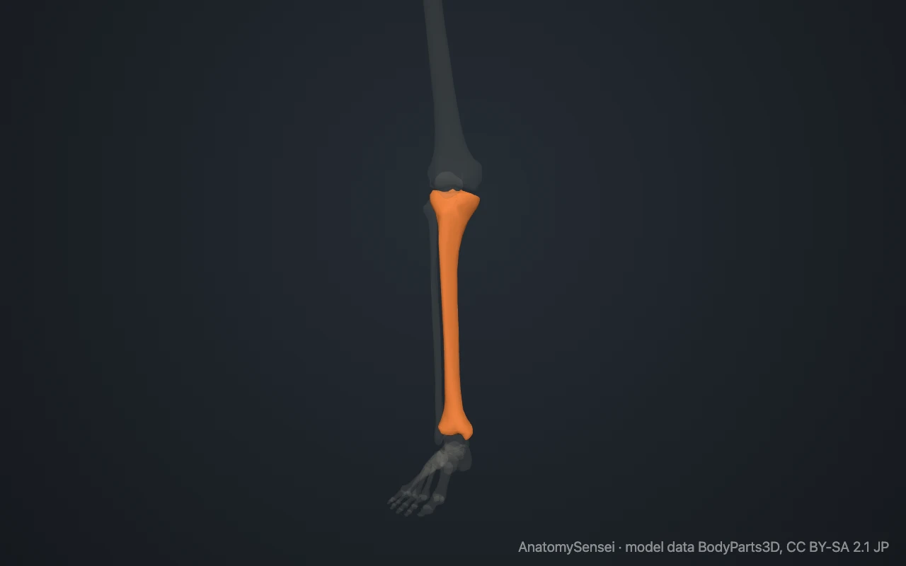
You can trace it with a finger. Start at the bump just below the kneecap, then follow the sharp ridge of the shin down the front of the leg. The flat surface to the inner side of that ridge is also tibia. At the bottom it ends in the bump on the inner side of the ankle.
What are the parts of the tibia?
The upper end is wide. It carries the medial and lateral condyles, whose flat tops together form the tibial plateau. Between them rises a small double peak, the intercondylar eminence.
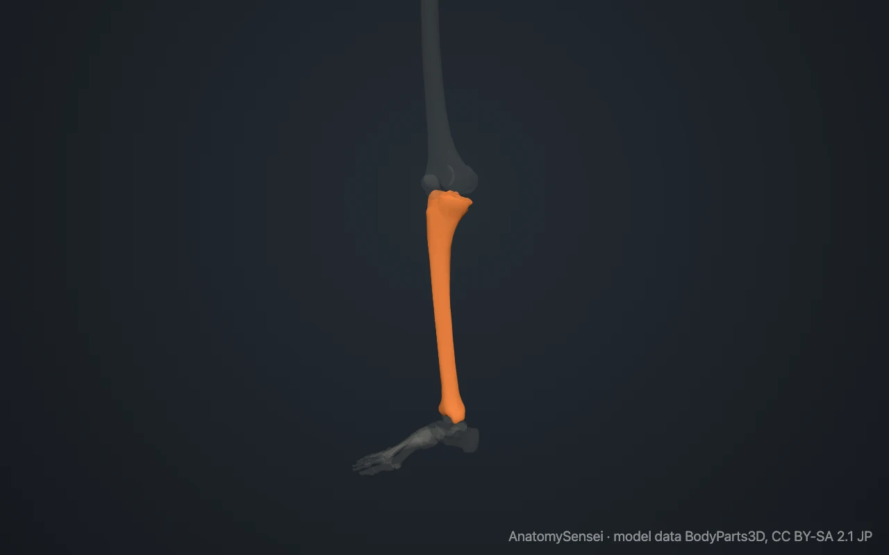
On the front, just below the condyles, is the tibial tuberosity. The shaft is triangular in cross-section. Its sharp anterior border is the shin, and its medial surface has no muscle covering it.
The lower end is smaller. On its inner side it extends downward as the medial malleolus. On its outer side is a shallow notch that receives the fibula.
What does the tibia articulate with?
The tibia meets three bones. At the knee, its two condyles support the condyles of the femur. At the ankle, its lower surface and the medial malleolus rest on the top and inner side of the talus.
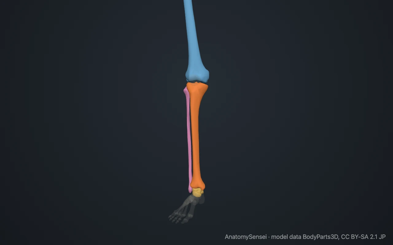
It meets the fibula twice. The head of the fibula touches the underside of the lateral condyle, and the lower end of the fibula fits into the notch above the ankle. The tibia does not form a joint with the patella.
What attaches to the tibia?
The patellar ligament attaches to the tibial tuberosity. Through it the quadriceps muscle pulls on the tibia to straighten the knee. On the top of the bone, the two menisci and the cruciate ligaments attach between and around the condyles.
Three thigh muscles, the sartorius, gracilis and semitendinosus, attach together on the upper inner surface. The tibialis anterior arises from the outer surface, and the soleus arises partly from a ridge on the back. A sheet of tissue, the interosseous membrane, ties the tibia to the fibula.
What does the tibia do?
It is the weight-bearing bone of the leg. Load from the femur spreads across the tibial plateau, travels down the shaft and passes to the talus.
It forms the lower half of the knee joint and the roof and inner wall of the ankle joint. It also gives origin to muscles that move the foot and toes.
Common problems with the tibia
The shaft of the tibia is one of the most commonly broken long bones. Because the bone lies just under the skin, a shaft fracture can break through it. A fracture of the upper end that enters the knee is called a tibial plateau fracture.
Pain along the inner edge of the shin after running is often called shin splints, and the tibia is a common site of stress fractures. In growing adolescents the tibial tuberosity can become sore and prominent, known as Osgood-Schlatter disease. This page describes anatomy only.
Common questions
Is the tibia the shin bone?
Yes. The sharp front edge of the tibia is the shin.
Which is bigger, the tibia or the fibula?
The tibia. It is much thicker and carries almost all of the weight.
Is the tibia on the inside or the outside of the leg?
On the inner side. The fibula is on the outer side.
Which bone forms the inner ankle bump?
The tibia. The bump is its lower end, the medial malleolus.
Is this 3D model of the tibia free?
Yes. The interactive 3D model on this page is free to view and use for study, with no account or download needed. Its geometry comes from the open BodyParts3D data set (CC BY-SA).
About this free 3D model of the tibia
The interactive 3D model on this page is free to use in your browser, with no sign-up. It is built from scan-based anatomy data, so every bone is a separate, correctly placed shape you can turn and inspect from any side.