
Femur (thigh bone)
Os femoris
The femur is the thigh bone. It is the longest and strongest bone in the body and runs from the hip to the knee.
Loading 3D model…
Free 3D model of the femur. Right leg. Drag to rotate, zoom with + and − (or Ctrl + scroll), click a bone to highlight and name it.
Key points
- The femur is the only bone in the thigh.
- Its rounded head fits into the socket of the hip bone to form the hip joint.
- Its two condyles rest on the tibia at the knee, and the patella glides on its front.
- It is the longest and strongest bone in the body.
What is the femur?
The femur, or thigh bone, is the only bone in the thigh. It is a long bone with a rounded upper end, a slightly bowed shaft and a broad lower end.
It is the longest and strongest bone in the body. It has to be, because it carries the whole weight of the trunk on each step and gives leverage to the largest muscles.
Where is the femur?
It runs from the hip to the knee, deep inside the muscles of the thigh. It does not hang straight down. The shaft slants inward, so the knees lie closer together than the hips.
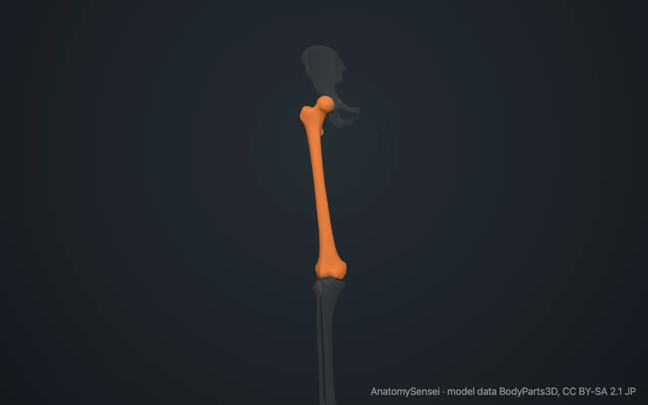
Most of the bone is too deep to feel, but its ends are not. Place a hand on the outer side of your hip, a hand width below the rim of the pelvis. The firm bump there is the greater trochanter. At the knee, the bony masses on each side just above the joint line are the lower ends of the femur.
What are the parts of the femur?
The upper end has a ball-shaped head that points upward and inward. It is joined to the shaft by the neck, which meets the shaft at an angle of about 125 degrees in adults.
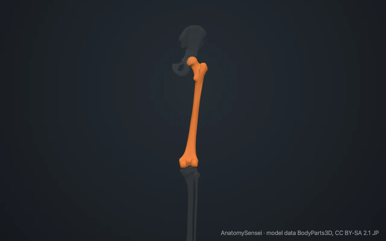
Where the neck meets the shaft there are two bumps for muscles. The greater trochanter is the large one on the outer side. The lesser trochanter is smaller and lies on the inner side at the back.
The shaft is smooth in front. Down its back runs a rough ridge called the linea aspera. The lower end widens into the medial and lateral condyles, two rounded knuckles separated at the back by a deep notch. Above each condyle is a small bump called an epicondyle.
What does the femur articulate with?
The femur meets three bones. At the hip joint its head sits in the acetabulum, the deep socket of the hip bone. This is a ball-and-socket joint.
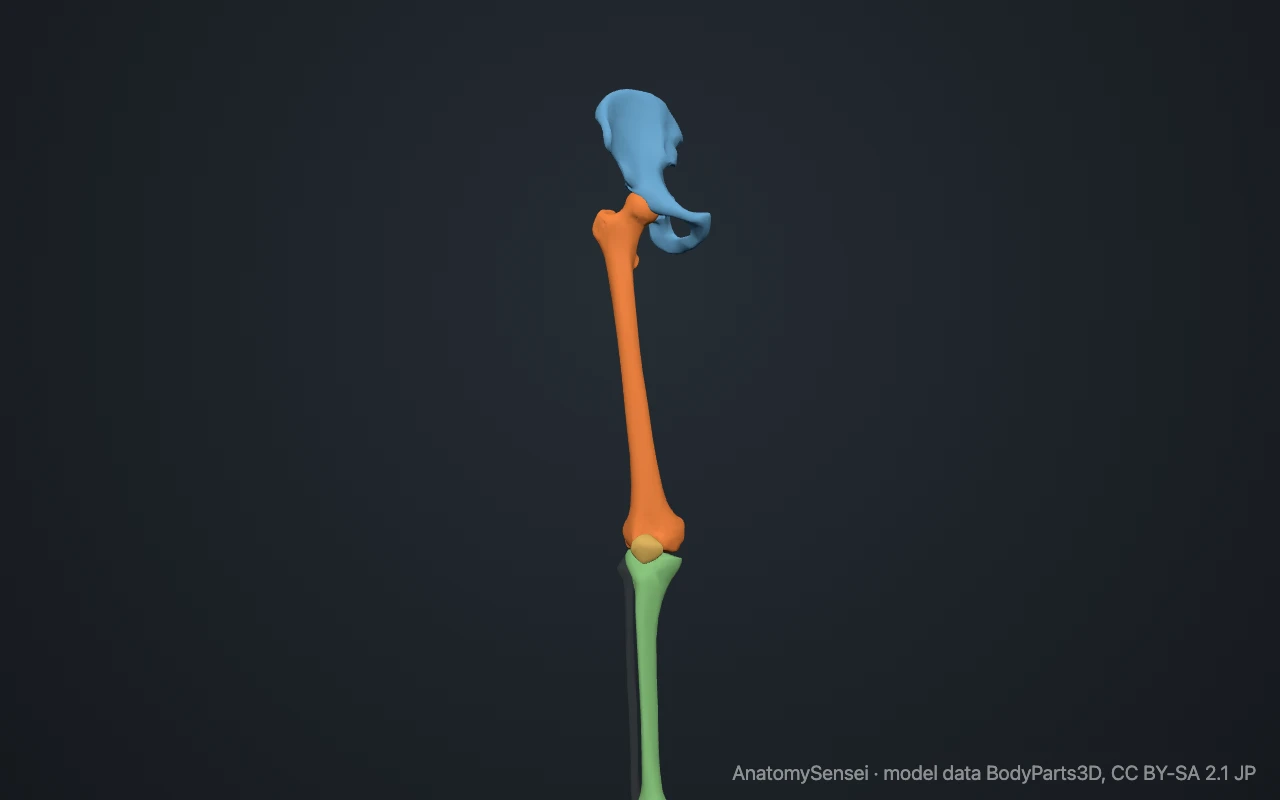
At the knee the two condyles rest on the top of the tibia. The patella glides in a groove on the front of the femur between the condyles. The femur does not touch the fibula.
What attaches to the femur?
The greater trochanter receives the gluteus medius and gluteus minimus, muscles that steady the pelvis during walking. The lesser trochanter receives the iliopsoas, the main muscle that bends the hip.
Three of the four parts of the quadriceps arise from the shaft. The adductor muscles of the inner thigh attach along the linea aspera. The two heads of the calf muscle, the gastrocnemius, arise from the back of the femur just above the condyles.
What does the femur do?
It carries body weight from the pelvis to the tibia. The angled neck holds the shaft away from the pelvis, which gives the hip a wide range of movement.
It also acts as a long lever for the muscles of the hip and knee. Its length is a large part of a person’s height.
Common problems with the femur
The neck of the femur is its weakest part. A break here or between the trochanters is what is usually meant by a hip fracture. It is most common in older people with thinner bones.
The shaft is very strong, and a shaft fracture usually needs a large force. The head can lose its blood supply, a condition called avascular necrosis. In adolescents the growth plate of the head can slip, known as slipped capital femoral epiphysis. This page describes anatomy only.
Common questions
Is the femur the longest bone in the body?
Yes. It is also the strongest.
Is the femur the same as the thigh bone?
Yes. Femur is the anatomical name for the thigh bone.
Can you feel your femur?
Only its ends. The greater trochanter can be felt on the outer side of the hip, and the condyles on each side of the knee.
Does the femur touch the fibula?
No. At the knee the femur meets only the tibia and the patella.
Is this 3D model of the femur free?
Yes. The interactive 3D model on this page is free to view and use for study, with no account or download needed. Its geometry comes from the open BodyParts3D data set (CC BY-SA).
About this free 3D model of the femur
The interactive 3D model on this page is free to use in your browser, with no sign-up. It is built from scan-based anatomy data, so every bone is a separate, correctly placed shape you can turn and inspect from any side.