
Pisiform bone
Os pisiforme
The pisiform is a pea-shaped bone at the base of the palm on the little finger side. It is the smallest carpal bone and a sesamoid bone, formed inside a tendon.
Loading 3D model…
Free 3D model of the pisiform bone. Right hand. Drag to rotate, zoom with + and − (or Ctrl + scroll), click a bone to highlight and name it.
Key points
- The pisiform is the smallest carpal bone, about the size of a pea.
- It sits on the palm surface of the triquetrum and joins no other bone.
- It is a sesamoid bone inside the tendon of flexor carpi ulnaris.
- It is easy to feel at the base of the palm on the little finger side.
What is the pisiform bone?
The pisiform is the smallest of the eight carpal bones. Its name comes from the Latin pisum, a pea, which describes both its size and its shape.
It is counted in the proximal row, but it is unlike the other carpals. It is a sesamoid bone, which means it forms inside a tendon. It takes no part in the wrist joint itself.
Where is the pisiform bone?
It lies at the base of the palm on the little finger side, in front of the triquetrum. It stands forward from the other carpals, so it shows clearly when the model is turned to the side.
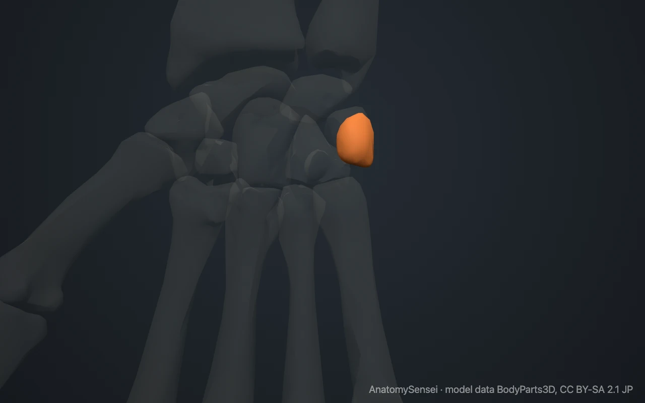
It is one of the easiest wrist bones to feel. Press on the heel of your hand on the little finger side, just beyond the wrist crease. The small hard knob there is the pisiform. With the wrist relaxed and slightly bent, it can be moved a little from side to side.
What does the pisiform look like?
The bone is small and rounded, like a pea or a small bean. Its back surface carries a single flat, oval facet for the triquetrum.
The rest of its surface is rough. The front faces the skin of the palm, and the sides give attachment to a tendon, a small muscle and several ligaments.
What does the pisiform articulate with?
The pisiform joins only one bone, the triquetrum. The small joint between them has its own capsule and lets the pisiform glide slightly as the wrist moves.
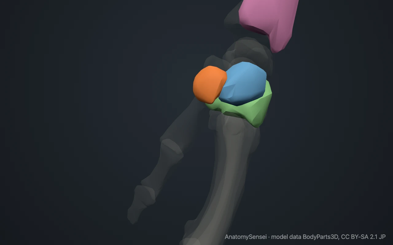
It does not touch the ulna, the hamate or any metacarpal. It is linked to the hamate and to the fifth metacarpal by ligaments instead.
What attaches to the pisiform?
The pisiform lies within the tendon of flexor carpi ulnaris, a forearm muscle that bends the wrist and tilts it towards the little finger. Two ligaments carry the pull of this tendon onward, one to the hook of the hamate and one to the base of the fifth metacarpal.
The abductor digiti minimi, the muscle that moves the little finger outward, starts from the pisiform. The flexor retinaculum, the roof of the carpal tunnel, is attached to it on the little finger side.
What passes beside the pisiform?
The ulnar nerve and ulnar artery enter the hand just to the thumb side of the pisiform. They run in a short passage between the pisiform and the hook of the hamate.
This passage is called the ulnar canal, or Guyon canal. The model shows its two bony walls, the pisiform and the hook of the hamate, but not the nerve or the artery.
What does the pisiform do?
Like other sesamoid bones, the pisiform works as a pulley. It holds the tendon of flexor carpi ulnaris away from the wrist, which improves the leverage of the muscle.
It also protects the tendon where it crosses the wrist and gives a firm starting point for the small muscle of the little finger.
Common problems with the pisiform
Pisiform fractures are uncommon. They usually follow a direct blow or a fall onto the heel of the hand. The joint between the pisiform and the triquetrum can also develop arthritis and become painful with gripping.
Pressure on the ulnar nerve in the canal beside the pisiform can cause numbness in the little finger and weakness of the hand. Long-distance cyclists who lean on the handlebars are a well-known example. This page describes anatomy only.
Common questions
Is the pisiform a sesamoid bone?
Yes. It forms inside the tendon of flexor carpi ulnaris.
Which bone does the pisiform join?
Only the triquetrum.
Can you feel the pisiform?
Yes. It is the hard knob at the base of the palm on the little finger side.
When does the pisiform form?
It is the last carpal bone to turn to bone, in later childhood. Before that it is cartilage and does not show on an X-ray.
Is this 3D model of the pisiform bone free?
Yes. The interactive 3D model on this page is free to view and use for study, with no account or download needed. Its geometry comes from the open BodyParts3D data set (CC BY-SA).
About this free 3D model of the pisiform bone
The interactive 3D model on this page is free to use in your browser, with no sign-up. It is built from scan-based anatomy data, so every bone is a separate, correctly placed shape you can turn and inspect from any side.