
Parts of the foot
Regiones pedis
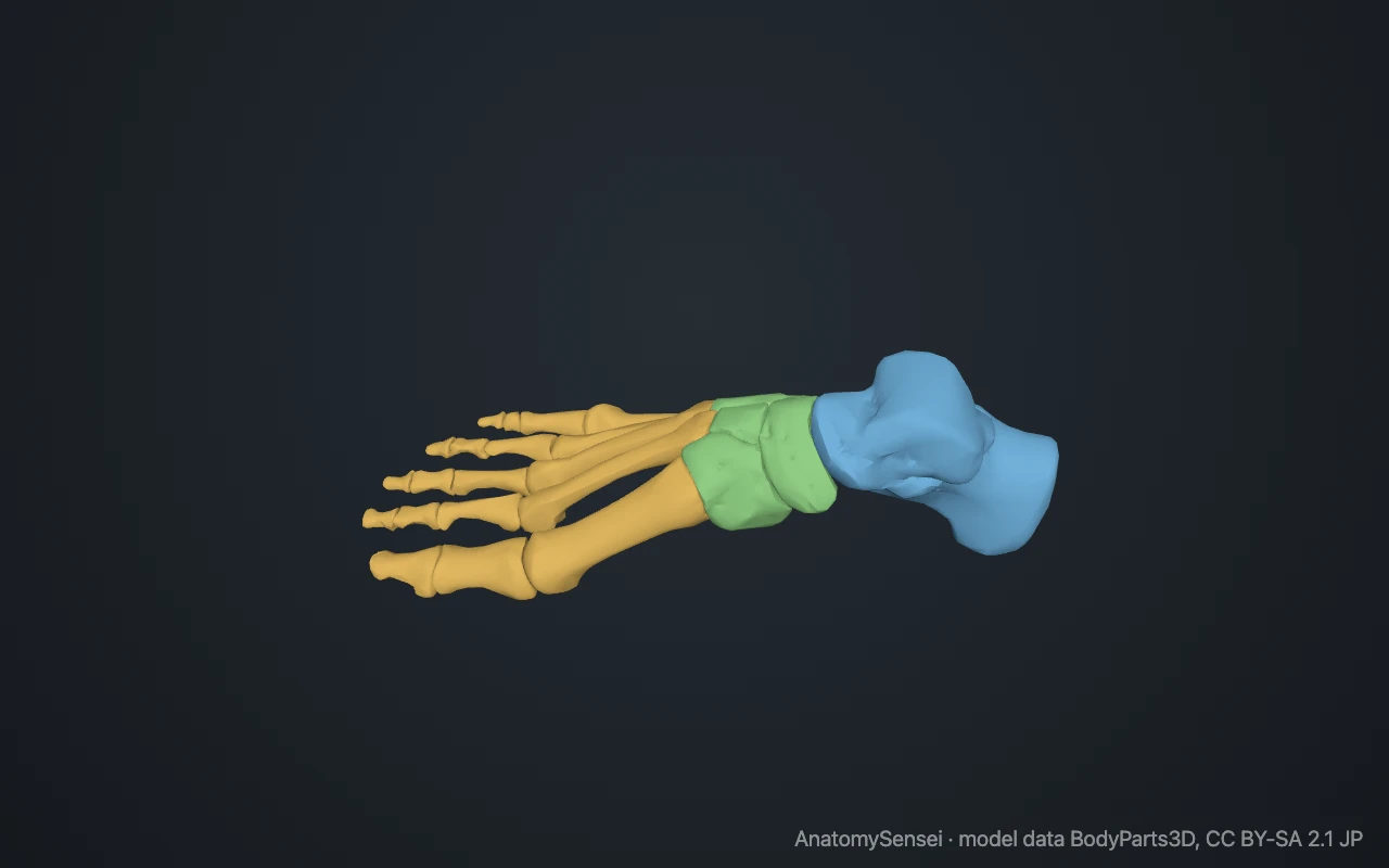
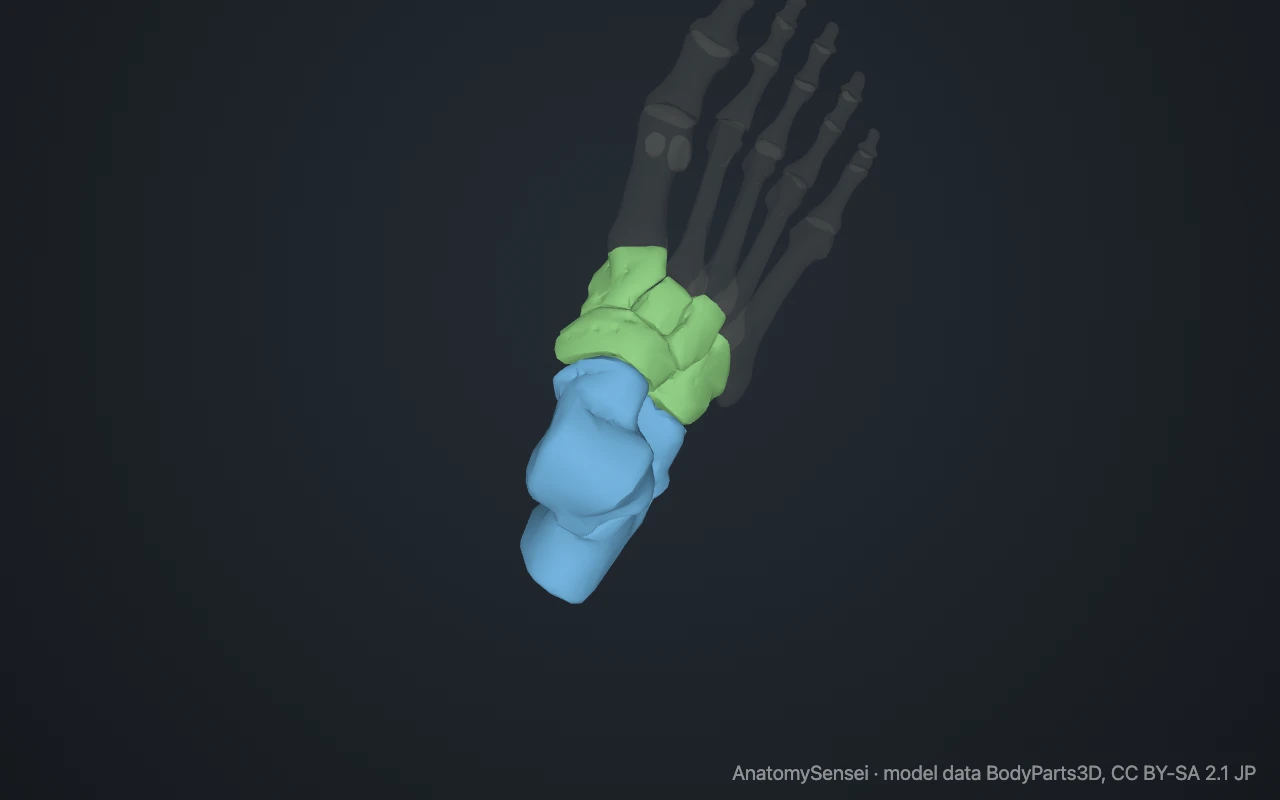
The foot has three parts. The hindfoot is the heel and ankle, the midfoot is the arch, and the forefoot is the ball of the foot and the toes.
Loading 3D model…
Free 3D model of the parts of the foot. Right foot. Drag to rotate, zoom with + and − (or Ctrl + scroll), click a bone to highlight and name it.
- Hindfoot: talus, calcaneus
- Midfoot: navicular, cuboid, cuneiforms
- Forefoot: metatarsals, phalanges, sesamoids
Key points
- The foot has three parts: hindfoot, midfoot and forefoot.
- The hindfoot is the heel and ankle; the midfoot is the arch; the forefoot is the ball and toes.
- The top of the foot is the dorsum; the underside is the sole, or plantar surface.
- Two joint lines separate the three parts.
What are the parts of the foot?
The foot is divided into three parts from back to front: the hindfoot, the midfoot and the forefoot. The division follows the bones and the joints between them.
The hindfoot
The hindfoot is the heel and the lower part of the ankle. It has two bones. The talus connects to the leg, and the calcaneus beneath it forms the heel.
These are the two largest bones in the foot. The hindfoot takes the impact when the foot lands and tilts in and out to keep you balanced.
The midfoot
The midfoot is the arch. It has five bones packed closely together: the navicular, the cuboid and the three cuneiforms.
It moves very little. Its job is to hold the arch and pass load from the hindfoot to the forefoot. The raised top of the midfoot is the instep.
The forefoot
The forefoot is the ball of the foot and the toes. It has the five metatarsals and the fourteen phalanges, plus the two sesamoids under the big toe.
It is the longest part and the last to leave the ground in each step.
Where one part ends and the next begins
Two joint lines mark the boundaries. The transverse tarsal joint, also called the Chopart joint, separates hindfoot from midfoot. The tarsometatarsal joints, also called the Lisfranc joint, separate midfoot from forefoot.
Top, sole and everyday names
The upper surface is the dorsum of the foot. The underside is the plantar surface, or sole. The inner border is medial and the outer border is lateral.
Everyday words fit the same map. The heel is the hindfoot. The arch and instep are the midfoot. The ball and the toes are the forefoot.
The inner and outer columns
The foot can also be read lengthwise. The inner column is the talus, navicular, cuneiforms and first three metatarsals. It is the higher, springier side.
The outer column is the calcaneus, cuboid and fourth and fifth metatarsals. It is lower and more mobile at the front, which lets the outer edge settle onto the ground.
Common questions
What is the forefoot?
The front part of the foot: the metatarsals and toes, including the ball of the foot.
What is the midfoot?
The middle part: the navicular, cuboid and three cuneiforms, which form the arch.
What is the hindfoot?
The back part: the talus and calcaneus.
What is the top of the foot called?
The dorsum. The underside is the sole, or plantar surface.
What is the ball of the foot?
The padded area behind the toes, over the heads of the metatarsals.
Is this 3D model of the parts of the foot free?
Yes. The interactive 3D model on this page is free to view and use for study, with no account or download needed. Its geometry comes from the open BodyParts3D data set (CC BY-SA).
About this free 3D model of the parts of the foot
The interactive 3D model on this page is free to use in your browser, with no sign-up. It is built from scan-based anatomy data, so every bone is a separate, correctly placed shape you can turn and inspect from any side.