
Sesamoid bones of the foot
Ossa sesamoidea pedis
A sesamoid bone is a small bone that grows inside a tendon. Each foot has two, about the size of a pea, under the head of the first metatarsal at the base of the big toe.
Loading 3D model…
Free 3D model of the sesamoid bones of the foot. Right foot. Drag to rotate, zoom with + and − (or Ctrl + scroll), click a bone to highlight and name it.
Key points
- A sesamoid is a small bone that forms inside a tendon.
- Each foot has two, side by side under the big toe joint, in the ball of the foot.
- They work as a pulley for the big toe and carry load when you push off.
- A sesamoid in two pieces is often a normal variant, not a fracture.
What is a sesamoid bone?
A sesamoid bone is a small bone that forms inside a tendon, at a point where the tendon bends around a joint. The name comes from the sesame seed, which the smallest ones resemble. Unlike other bones, a sesamoid is not joined to its neighbours by a joint capsule of its own. It rides in the tendon and glides over the bone beneath it.
The kneecap is the largest sesamoid in the body. The two under the big toe are the next most important, because they carry body weight with every step.
Where are the sesamoid bones in the foot?
They sit in the ball of the foot, directly under the head of the first metatarsal, which is the long bone behind the big toe. Each is about the size of a pea or a small bean.
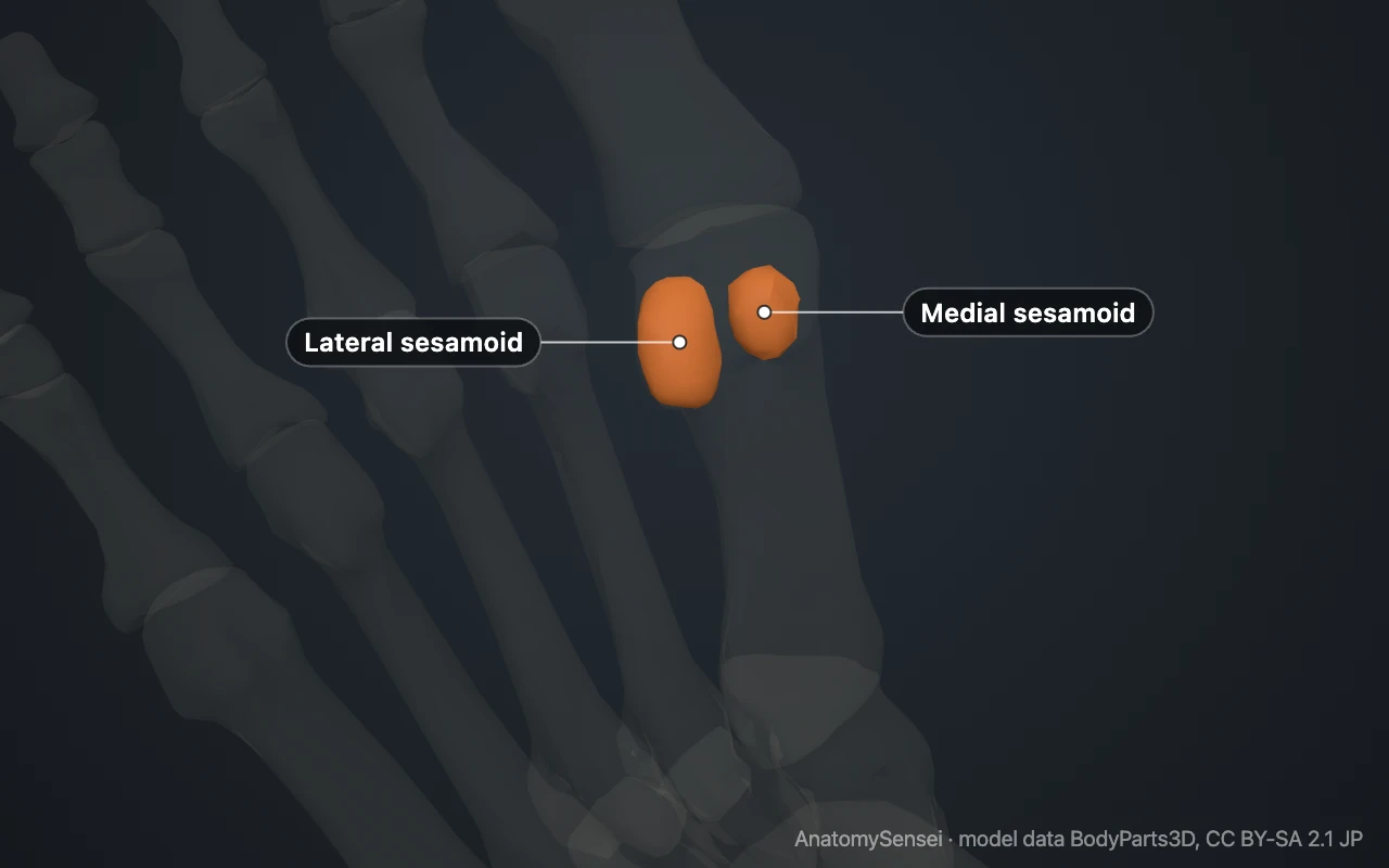
You can find the spot on your own foot. Press into the sole just behind the big toe, at the thickest part of the ball of the foot. The firm lump under the skin is the head of the first metatarsal with the two sesamoids beneath it. They cannot be seen from above, which is why the model on this page opens from the sole.
The medial and lateral sesamoid
The two bones lie side by side. The medial sesamoid is on the inner side of the foot, toward the other foot, and is also called the tibial sesamoid. The lateral sesamoid is on the outer side and is also called the fibular sesamoid.
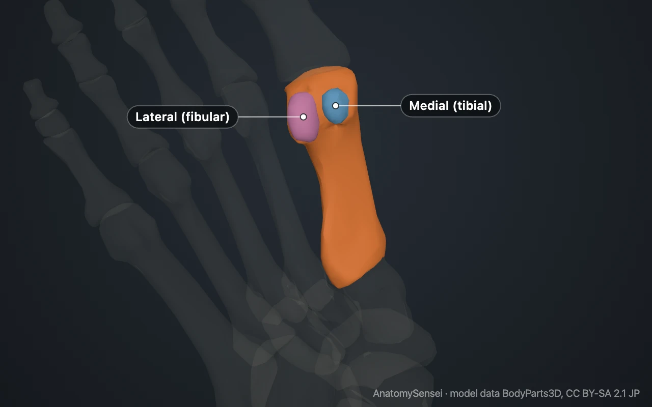
Each one sits in its own shallow groove on the underside of the metatarsal head. A low ridge of bone between the grooves keeps them on track, and a short ligament ties the two together. The medial sesamoid is usually the larger and takes more of the load.
What do the sesamoid bones do?
They do three jobs. First, they act as a pulley. By holding the tendons a little away from the joint, they give the muscle that bends the big toe more leverage, so the toe can push harder against the ground.
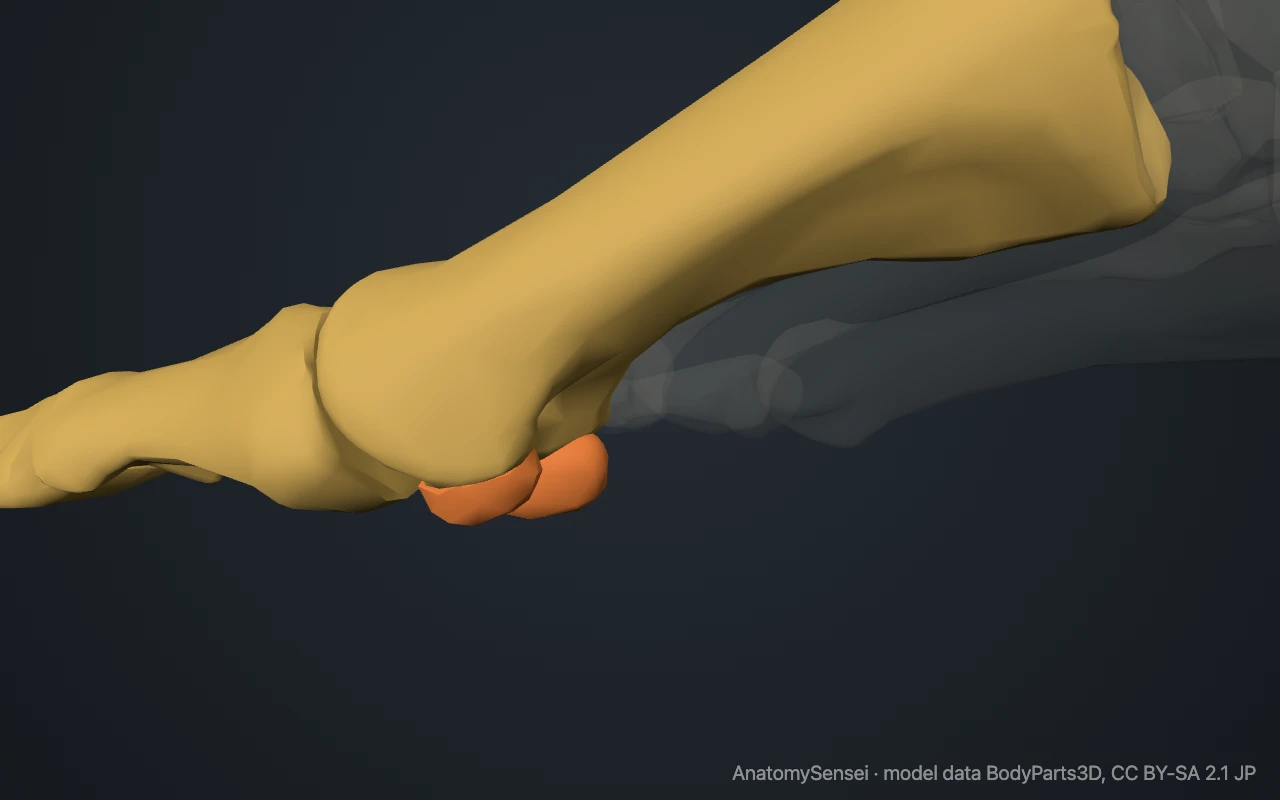
Second, they bear weight. When the heel lifts and the foot rolls forward, load passes through the sesamoids before the toe leaves the ground. Third, they protect the long tendon that runs to the tip of the big toe, which passes through the channel between them and would otherwise be pressed against the ground.
What attaches to them?
Both sesamoids are embedded in the two tendons of flexor hallucis brevis, the short muscle that bends the big toe. The abductor hallucis muscle joins the medial sesamoid, and the adductor hallucis joins the lateral one. In front, the sesamoids are anchored to the base of the first toe bone through a thick pad of tissue called the plantar plate.
The tendon of flexor hallucis longus does not attach to them. It runs forward in the gap between the two bones.
Are there other sesamoid bones in the foot?
Sometimes. The pair under the big toe is present in almost everyone. Extra sesamoids can also occur at the joint in the middle of the big toe and under the heads of the other metatarsals, most often the second and the fifth. A separate one, the os peroneum, may form in a tendon on the outer side of the foot next to the cuboid bone.
These extra bones vary from person to person and are usually found by chance on an X-ray. The model here shows only the two that are reliably present.
Common problems with the sesamoids
Because they carry load in a small area, the sesamoids are prone to overuse. Sesamoiditis is pain and irritation around them, typical in runners and dancers. They can also fracture, either from a hard landing or gradually as a stress fracture. Turf toe is a sprain of the tissue that holds them in place, caused by the big toe being forced upward.
One feature is often mistaken for a break. In some people the medial sesamoid develops in two pieces, called a bipartite sesamoid. It is a normal variant and often appears in both feet. Lasting pain under the big toe joint is a reason to see a clinician; this page describes the anatomy only.
Common questions
How many sesamoid bones are in the foot?
Two that are almost always present, both under the head of the first metatarsal. Some people have additional small ones elsewhere in the foot.
Can you feel the sesamoid bones?
Not as two separate bones, but you can feel where they are: the firm area in the ball of the foot just behind the big toe.
Which sesamoid is bigger, medial or lateral?
Usually the medial one. It also takes more load, which is one reason it is the one more often injured.
Is a sesamoid in two pieces always a fracture?
No. A bipartite sesamoid is a normal variant in which the bone formed in two parts. Telling it apart from a fracture is done on imaging.
How many sesamoid bones are in the body?
There is no fixed number. The two kneecaps and the pair under each big toe are constant, the thumb usually has two, and others vary between people.
Is this 3D model of the sesamoid bones of the foot free?
Yes. The interactive 3D model on this page is free to view and use for study, with no account or download needed. Its geometry comes from the open BodyParts3D data set (CC BY-SA).
About this free 3D model of the sesamoid bones of the foot
The interactive 3D model on this page is free to use in your browser, with no sign-up. It is built from scan-based anatomy data, so every bone is a separate, correctly placed shape you can turn and inspect from any side.